Yeh Connie Y, Koehl Kristin L, Harman Christine D, Iwabe Simone, Guzman José M, Petersen-Jones Simon M, Kardon Randy H, Komáromy András M
College of Veterinary Medicine, Michigan State University, East Lansing, Michigan, United States 2School of Veterinary Medicine, University of Pennsylvania, Philadelphia, Pennsylvania, United States.
College of Veterinary Medicine, Michigan State University, East Lansing, Michigan, United States.
Invest Ophthalmol Vis Sci. 2017 Jan 1;58(1):65-78. doi: 10.1167/iovs.16-19865.
The purpose of this study was to evaluate a chromatic pupillometry protocol for specific functional assessment of rods, cones, and intrinsically photosensitive retinal ganglion cells (ipRGCs) in dogs.
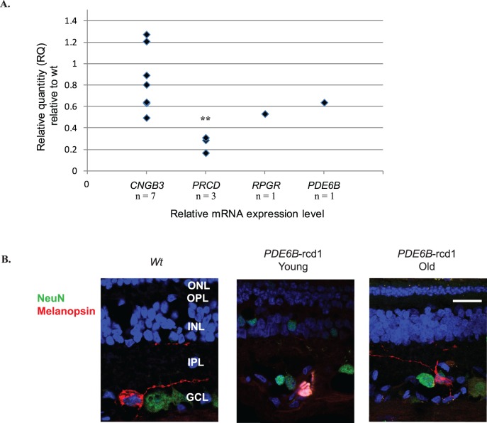
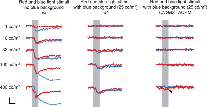
Chromatic pupillometry was tested and compared in 37 dogs in different stages of primary loss of rod, cone, and combined rod/cone and optic nerve function, and in 5 wild-type (WT) dogs. Eyes were stimulated with 1-s flashes of dim (1 cd/m2) and bright (400 cd/m2) blue light (for scotopic conditions) or bright red (400 cd/m2) light with 25-cd/m2 blue background (for photopic conditions). Canine retinal melanopsin/Opn4 was cloned, and its expression was evaluated using real-time quantitative reverse transcription-PCR and immunohistochemistry.
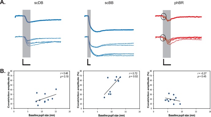
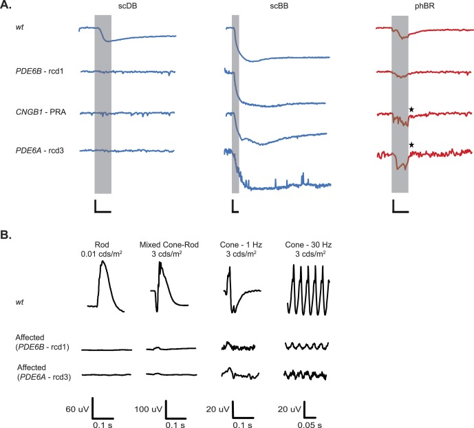
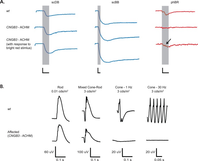
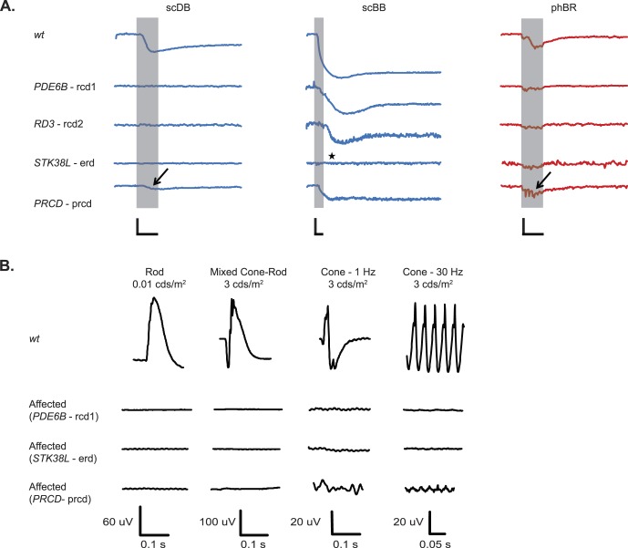
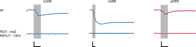
Mean ± SD percentage of pupil constriction amplitudes induced by scotopic dim blue (scDB), scotopic bright blue (scBB), and photopic bright red (phBR) lights in WT dogs were 21.3% ± 10.6%, 50.0% ± 17.5%, and 19.4% ± 7.4%, respectively. Melanopsin-mediated responses to scBB persisted for several minutes (7.7 ± 4.6 min) after stimulus offset. In dogs with inherited retinal degeneration, loss of rod function resulted in absent scDB responses, followed by decreased phBR responses with disease progression and loss of cone function. Primary loss of cone function abolished phBR responses but preserved those responses to blue light (scDB and scBB). Although melanopsin/Opn4 expression was diminished with retinal degeneration, melanopsin-expressing ipRGCs were identified for the first time in both WT and degenerated canine retinas.
Pupil responses elicited by light stimuli of different colors and intensities allowed differential functional assessment of canine rods, cones, and ipRGCs. Chromatic pupillometry offers an effective tool for diagnosing retinal and optic nerve diseases.
本研究旨在评估一种用于犬视杆细胞、视锥细胞和内在光敏性视网膜神经节细胞(ipRGCs)特定功能评估的彩色瞳孔测量方案。
对37只处于视杆细胞、视锥细胞、视杆/视锥细胞联合及视神经功能原发性丧失不同阶段的犬以及5只野生型(WT)犬进行彩色瞳孔测量测试并比较。眼睛分别用1秒的昏暗(1 cd/m²)和明亮(400 cd/m²)蓝光闪烁(用于暗视条件)或带有25 cd/m²蓝色背景的明亮红色(400 cd/m²)光闪烁(用于明视条件)进行刺激。克隆犬视网膜黑视蛋白/Opn4,并使用实时定量逆转录聚合酶链反应和免疫组织化学评估其表达。
野生型犬中,暗视昏暗蓝光(scDB)、暗视明亮蓝光(scBB)和明视明亮红光(phBR)刺激引起的平均瞳孔收缩幅度百分比±标准差分别为21.3%±10.6%、50.0%±17.5%和19.4%±7.4%。黑视蛋白介导的对scBB的反应在刺激停止后持续数分钟(7.7±4.6分钟)。在患有遗传性视网膜变性的犬中,视杆细胞功能丧失导致scDB反应消失,随后随着疾病进展和视锥细胞功能丧失,phBR反应降低。视锥细胞功能原发性丧失消除了phBR反应,但保留了对蓝光的反应(scDB和scBB)。尽管随着视网膜变性黑视蛋白/Opn4表达减少,但首次在野生型和变性犬视网膜中均鉴定出表达黑视蛋白的ipRGCs。
不同颜色和强度光刺激引起的瞳孔反应允许对犬视杆细胞、视锥细胞和ipRGCs进行差异性功能评估。彩色瞳孔测量为诊断视网膜和视神经疾病提供了一种有效工具。