• 文献检索
  • 文档翻译
  • 深度研究
  • 学术资讯
  • Suppr Zotero 插件Zotero 插件
  • 邀请有礼
  • 套餐&价格
  • 历史记录
应用&插件
Suppr Zotero 插件Zotero 插件浏览器插件Mac 客户端Windows 客户端微信小程序
定价
高级版会员购买积分包购买API积分包
服务
文献检索文档翻译深度研究API 文档MCP 服务
关于我们
关于 Suppr公司介绍联系我们用户协议隐私条款
关注我们

Suppr 超能文献

核心技术专利:CN118964589B侵权必究
粤ICP备2023148730 号-1Suppr @ 2026

文献检索

告别复杂PubMed语法,用中文像聊天一样搜索,搜遍4000万医学文献。AI智能推荐,让科研检索更轻松。

立即免费搜索

文件翻译

保留排版,准确专业,支持PDF/Word/PPT等文件格式,支持 12+语言互译。

免费翻译文档

深度研究

AI帮你快速写综述,25分钟生成高质量综述,智能提取关键信息,辅助科研写作。

立即免费体验

用于摄取研究的细胞外囊泡亲脂性荧光团染色相关的陷阱。

Pitfalls associated with lipophilic fluorophore staining of extracellular vesicles for uptake studies.

作者信息

Simonsen Jens B

机构信息

Department of Health Technology (DTU Health Tech) and Department of Micro- and Nanotechnology (DTU Nanotech), Technical University of Denmark, Lyngby, Denmark.

出版信息

J Extracell Vesicles. 2019 Feb 20;8(1):1582237. doi: 10.1080/20013078.2019.1582237. eCollection 2019.

DOI:10.1080/20013078.2019.1582237
PMID:30815239
原文链接:https://pmc.ncbi.nlm.nih.gov/articles/PMC6383605/
Abstract

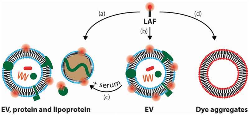
Post-staining of extracellular vesicles (EVs) with lipid-anchored fluorophores (LAFs) such as PKH67 is a widely used strategy for studying EVs but it is associated with several pitfalls. The pitfalls discussed in this commentary are related to LAF labelling of non-EV species due to (1) lipoprotein contamination in EV samples, (2) desorption of the LAF reporters from vesicles into proteins and lipoproteins in blood and serum, and (3) the capability of the amphiphilic LAF compounds to form EV-like particles. Awareness of these challenges and developing solutions to overcome these are important to ensure that we make relevant interpretations when using LAFs to track EVs.

摘要

使用脂质锚定荧光团(LAF)(如PKH67)对细胞外囊泡(EV)进行染色后处理是研究EV的一种广泛使用的策略,但它存在几个缺陷。本评论中讨论的缺陷与非EV物种的LAF标记有关,原因如下:(1)EV样本中的脂蛋白污染;(2)LAF报告分子从囊泡解吸到血液和血清中的蛋白质和脂蛋白中;(3)两亲性LAF化合物形成类似EV颗粒的能力。认识到这些挑战并开发克服这些挑战的解决方案,对于确保我们在使用LAF追踪EV时做出相关解释非常重要。

https://cdn.ncbi.nlm.nih.gov/pmc/blobs/8fbf/6383605/9e56b97dfcd0/ZJEV_A_1582237_F0001_OC.jpg

相似文献

1
Pitfalls associated with lipophilic fluorophore staining of extracellular vesicles for uptake studies.用于摄取研究的细胞外囊泡亲脂性荧光团染色相关的陷阱。
J Extracell Vesicles. 2019 Feb 20;8(1):1582237. doi: 10.1080/20013078.2019.1582237. eCollection 2019.
2
Quantitative assessment of lipophilic membrane dye-based labelling of extracellular vesicles by nano-flow cytometry.纳米流式细胞术定量评估基于亲脂性膜染料的细胞外囊泡标记。
J Extracell Vesicles. 2023 Aug;12(8):e12351. doi: 10.1002/jev2.12351.
3
Fluorescent Labeling of Helminth Extracellular Vesicles Using an In Vivo Whole Organism Approach.使用体内全生物体方法对蠕虫细胞外囊泡进行荧光标记
Biomedicines. 2020 Jul 14;8(7):213. doi: 10.3390/biomedicines8070213.
4
Specific and Non-Invasive Fluorescent Labelling of Extracellular Vesicles for Evaluation of Intracellular Processing by Intestinal Epithelial Cells.用于评估肠道上皮细胞细胞内加工过程的细胞外囊泡特异性非侵入性荧光标记
Biomedicines. 2020 Jul 14;8(7):211. doi: 10.3390/biomedicines8070211.
5
Lipid Profile Characterization and Lipoprotein Comparison of Extracellular Vesicles from Human Plasma and Serum.人血浆和血清来源细胞外囊泡的脂质谱特征及脂蛋白比较
Metabolites. 2019 Nov 1;9(11):259. doi: 10.3390/metabo9110259.
6
Refractive index to evaluate staining specificity of extracellular vesicles by flow cytometry.通过流式细胞术评估细胞外囊泡染色特异性的折射率。
J Extracell Vesicles. 2019 Jul 25;8(1):1643671. doi: 10.1080/20013078.2019.1643671. eCollection 2019.
7
Isolation and Characterization of Functionally Active Extracellular Vesicles from Culture Medium Conditioned by Bovine Embryos In Vitro.从体外培养的牛胚胎条件培养基中分离和表征功能活跃的细胞外囊泡。
Int J Mol Sci. 2018 Dec 21;20(1):38. doi: 10.3390/ijms20010038.
8
Isolation of High-Purity Extracellular Vesicles by the Combination of Iodixanol Density Gradient Ultracentrifugation and Bind-Elute Chromatography From Blood Plasma.通过碘克沙醇密度梯度超速离心和结合洗脱色谱法从血浆中分离高纯度细胞外囊泡。
Front Physiol. 2018 Oct 23;9:1479. doi: 10.3389/fphys.2018.01479. eCollection 2018.
9
Analyzing the miRNA content of extracellular vesicles by fluorescence nanoparticle tracking.通过荧光纳米颗粒追踪分析细胞外囊泡的微小RNA含量。
Nanomedicine. 2017 Feb;13(2):765-770. doi: 10.1016/j.nano.2016.10.013. Epub 2016 Oct 28.
10
Systematic characterization of extracellular vesicle sorting domains and quantification at the single molecule - single vesicle level by fluorescence correlation spectroscopy and single particle imaging.通过荧光相关光谱和单粒子成像对细胞外囊泡分选结构域进行系统表征及在单分子-单囊泡水平上进行定量分析。
J Extracell Vesicles. 2019 Sep 18;8(1):1663043. doi: 10.1080/20013078.2019.1663043. eCollection 2019.

引用本文的文献

1
Ontogenetic stage and type of donor cells shape extracellular vesicles' therapeutic potential for osteoarthritis.个体发育阶段和供体细胞类型塑造了细胞外囊泡对骨关节炎的治疗潜力。
Stem Cell Res Ther. 2025 Sep 1;16(1):478. doi: 10.1186/s13287-025-04585-y.
2
Roadblocks of Urinary EV Biomarkers: Moving Toward the Clinic.尿液细胞外囊泡生物标志物的障碍:迈向临床应用
J Extracell Vesicles. 2025 Jul;14(7):e70120. doi: 10.1002/jev2.70120.
3
Uptake of DU145 and LNCaP prostate cancer cell line derived extracellular vesicles is inversely correlated with blood-brain barrier integrity in vitro.

本文引用的文献

1
What is the blood concentration of extracellular vesicles? Implications for the use of extracellular vesicles as blood-borne biomarkers of cancer.细胞外囊泡的血浓度是多少?这对将细胞外囊泡作为癌症血源性生物标志物的应用有何影响。
Biochim Biophys Acta Rev Cancer. 2019 Jan;1871(1):109-116. doi: 10.1016/j.bbcan.2018.11.006. Epub 2018 Dec 7.
2
Dissociation of fluorescently labeled lipids from liposomes in biological environments challenges the interpretation of uptake studies.荧光标记脂质从脂质体中在生物环境中的解离,给摄取研究的解释带来了挑战。
Nanoscale. 2018 Dec 13;10(48):22720-22724. doi: 10.1039/c8nr07755j.
3
Circulating Extracellular Vesicles in Human Disease.
DU145和LNCaP前列腺癌细胞系来源的细胞外囊泡的摄取在体外与血脑屏障完整性呈负相关。
Fluids Barriers CNS. 2025 Jul 7;22(1):70. doi: 10.1186/s12987-025-00680-7.
4
Clinical Scale MSC-Derived Extracellular Vesicles Enhance Poststroke Neuroplasticity in Rodents and Non-Human Primates.临床级间充质干细胞衍生的细胞外囊泡增强啮齿动物和非人类灵长类动物中风后的神经可塑性。
J Extracell Vesicles. 2025 Jun;14(6):e70110. doi: 10.1002/jev2.70110.
5
Benzimidazole-Derived B2 as a Fluorescent Probe for Bacterial Outer Membrane Vesicle (OMV) Labeling: Integrating DFT, Molecular Dynamics, Flow Cytometry, and Confocal Microscopy.作为用于细菌外膜囊泡(OMV)标记的荧光探针的苯并咪唑衍生物B2:整合密度泛函理论、分子动力学、流式细胞术和共聚焦显微镜技术
Int J Mol Sci. 2025 May 14;26(10):4682. doi: 10.3390/ijms26104682.
6
Non-Specific Particle Formation During Extracellular Vesicle Labelling With the Lipophilic Membrane Dye PKH26.使用亲脂性膜染料PKH26标记细胞外囊泡期间的非特异性颗粒形成
J Extracell Vesicles. 2025 May;14(5):e70079. doi: 10.1002/jev2.70079.
7
The INAVA mRNA in Extracellular Vesicles Activates Normal Ovarian Fibroblasts by Phosphorylation-Ubiquitylation Crosstalk of HMGA2.细胞外囊泡中的INAVA mRNA通过HMGA2的磷酸化-泛素化串扰激活正常卵巢成纤维细胞。
Adv Sci (Weinh). 2025 Jul;12(25):e2500912. doi: 10.1002/advs.202500912. Epub 2025 Apr 23.
8
Advances in Tracing Techniques: Mapping the Trajectory of Mesenchymal Stem-Cell-Derived Extracellular Vesicles.追踪技术的进展:绘制间充质干细胞衍生细胞外囊泡的轨迹
Chem Biomed Imaging. 2025 Feb 2;3(3):137-168. doi: 10.1021/cbmi.4c00085. eCollection 2025 Mar 24.
9
Extracellular Vesicles Derived From Regenerating Tissue Promote Stem Cell Proliferation in the Planarian .源自再生组织的细胞外囊泡促进涡虫体内干细胞增殖
J Extracell Biol. 2025 Mar 9;4(3):e70040. doi: 10.1002/jex2.70040. eCollection 2025 Mar.
10
Reply to: Letter to Editor by Saravanan Sampoornam et al.回复:萨拉瓦南·桑普尔南姆等人致编辑的信
J Cell Mol Med. 2025 Mar;29(5):e70465. doi: 10.1111/jcmm.70465.
人类疾病中的循环细胞外囊泡
N Engl J Med. 2018 Sep 6;379(10):958-966. doi: 10.1056/NEJMra1704286.
4
PKH26 labeling of extracellular vesicles: Characterization and cellular internalization of contaminating PKH26 nanoparticles.PKH26 标记细胞外囊泡:污染的 PKH26 纳米颗粒的表征和细胞内化。
Biochim Biophys Acta Biomembr. 2018 Jun;1860(6):1350-1361. doi: 10.1016/j.bbamem.2018.03.013. Epub 2018 Mar 16.
5
Detailed analysis of the plasma extracellular vesicle proteome after separation from lipoproteins.详细分析脂蛋白分离后的血浆细胞外囊泡蛋白质组。
Cell Mol Life Sci. 2018 Aug;75(15):2873-2886. doi: 10.1007/s00018-018-2773-4. Epub 2018 Feb 13.
6
Shedding light on the cell biology of extracellular vesicles.揭示细胞外囊泡的细胞生物学。
Nat Rev Mol Cell Biol. 2018 Apr;19(4):213-228. doi: 10.1038/nrm.2017.125. Epub 2018 Jan 17.
7
Confounding factors in vesicle uptake studies using fluorescent lipophilic membrane dyes.使用荧光亲脂性膜染料进行囊泡摄取研究中的混杂因素。
J Extracell Vesicles. 2017 Oct 12;6(1):1388731. doi: 10.1080/20013078.2017.1388731. eCollection 2017.
8
On the use of liposome controls in studies investigating the clinical potential of extracellular vesicle-based drug delivery systems - A commentary.关于在研究基于细胞外囊泡的药物递送系统的临床潜力的研究中使用脂质体对照的评论。
J Control Release. 2018 Jan 10;269:10-14. doi: 10.1016/j.jconrel.2017.11.002. Epub 2017 Nov 7.
9
What Are We Looking At? Extracellular Vesicles, Lipoproteins, or Both?我们在看什么?细胞外囊泡、脂蛋白,还是两者皆有?
Circ Res. 2017 Sep 29;121(8):920-922. doi: 10.1161/CIRCRESAHA.117.311767.
10
Achieving the Promise of Therapeutic Extracellular Vesicles: The Devil is in Details of Therapeutic Loading.实现治疗性细胞外囊泡的前景:关键在于治疗性负载的细节。
Pharm Res. 2017 May;34(5):1053-1066. doi: 10.1007/s11095-017-2123-5. Epub 2017 Mar 17.