• 文献检索
  • 文档翻译
  • 深度研究
  • 学术资讯
  • Suppr Zotero 插件Zotero 插件
  • 邀请有礼
  • 套餐&价格
  • 历史记录
应用&插件
Suppr Zotero 插件Zotero 插件浏览器插件Mac 客户端Windows 客户端微信小程序
定价
高级版会员购买积分包购买API积分包
服务
文献检索文档翻译深度研究API 文档MCP 服务
关于我们
关于 Suppr公司介绍联系我们用户协议隐私条款
关注我们

Suppr 超能文献

核心技术专利:CN118964589B侵权必究
粤ICP备2023148730 号-1Suppr @ 2026

文献检索

告别复杂PubMed语法,用中文像聊天一样搜索,搜遍4000万医学文献。AI智能推荐,让科研检索更轻松。

立即免费搜索

文件翻译

保留排版,准确专业,支持PDF/Word/PPT等文件格式,支持 12+语言互译。

免费翻译文档

深度研究

AI帮你快速写综述,25分钟生成高质量综述,智能提取关键信息,辅助科研写作。

立即免费体验

内镜可见光谱评估:在猪模型中与微血管氧张力测量的比较。

Evaluation of endoscopic visible light spectroscopy: comparison with microvascular oxygen tension measurements in a porcine model.

机构信息

Department of Anesthesiology, Laboratory for Experimental Anesthesiology, Erasmus MC University Medical Center, s Gravendijkwal 230, 3015 CE, Rotterdam, The Netherlands.

Department of Gastroenterology and Hepatology, Erasmus MC University Medical Center, s Gravendijkwal 230, 3015 CE, Rotterdam, The Netherlands.

出版信息

J Transl Med. 2019 Feb 28;17(1):65. doi: 10.1186/s12967-019-1802-x.

DOI:10.1186/s12967-019-1802-x
PMID:30819196
原文链接:https://pmc.ncbi.nlm.nih.gov/articles/PMC6396526/
Abstract

BACKGROUND

Visible light spectroscopy (VLS) is a technique used to measure the mucosal oxygen saturation during upper gastrointestinal endoscopy to evaluate mucosal ischemia, however in vivo validation is lacking. We aimed to compare VLS measurements with a validated quantitative microvascular oxygen tension (μPO) measurement technique.

METHODS

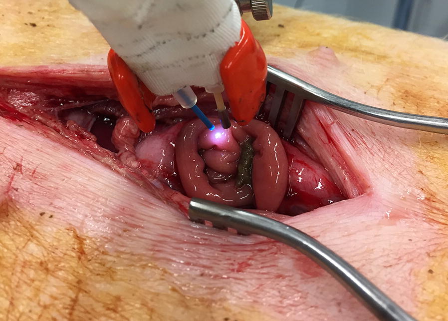
Simultaneous VLS measurements and μPO measurements were performed on the small intestine of five pigs. First, simultaneous measurements were performed at different FiO values (18%-100%). Thereafter, the influence of bile was assessed by comparing VLS measurements in the presence of bile and without bile. Finally, simultaneous VLS and μPO measurements were performed from the moment a lethal dose potassium chloride intravenously was injected.

RESULTS

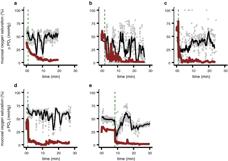
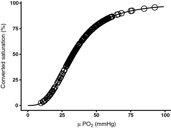
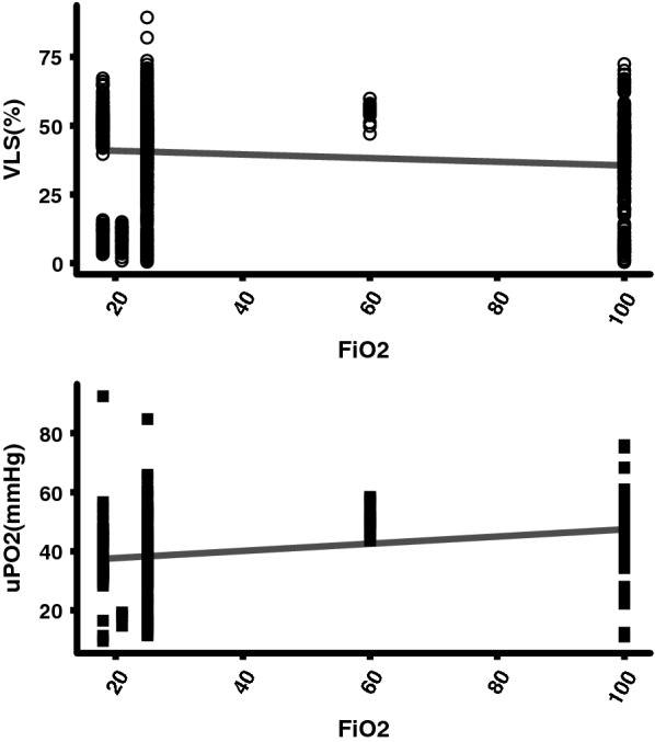
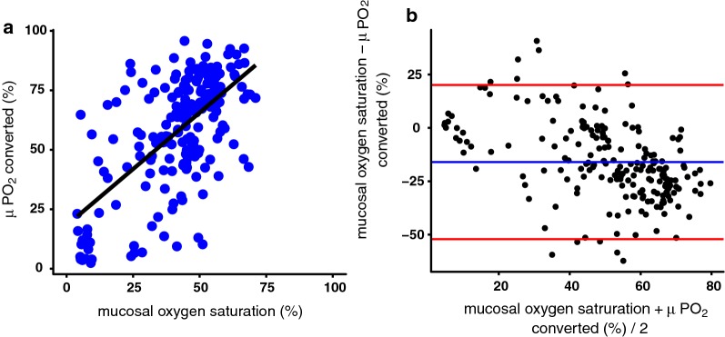
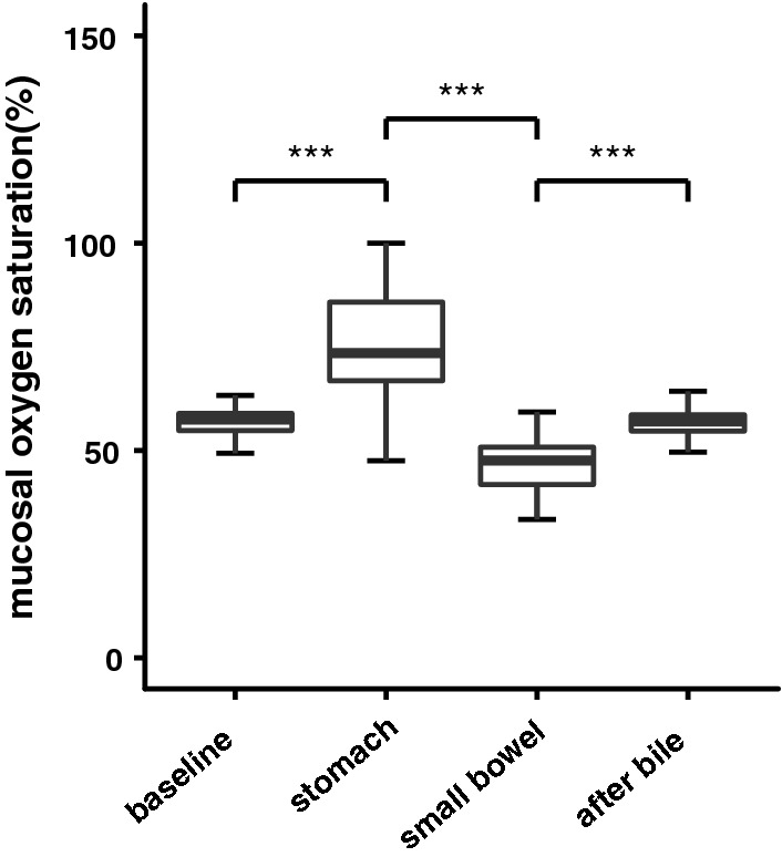
In contrast to μPO values that increased with increasing FiO, VLS values decreased. Both measurements correlated poorly with R = 0.39, intercept 18.5, slope 0.41 and a bias of - 16%. Furthermore, the presence of bile influenced VLS values significantly (median (IQR)) before bile application 57.5% (54.8-59.0%) versus median with bile mixture of the stomach 73.5% (66.8-85.8), p = < 2.2 * 10; median with bile mixture of small bowel 47.6% (41.8-50.8) versus median after bile removal 57.0% (54.7-58.6%), p = < 2.2 * 10). Finally, the VLS mucosal oxygen saturation values did not decrease towards a value of 0 in the first 25 min of asystole in contrast to the μPO values.

CONCLUSIONS

These results suggest that VLS measures the mixed venous oxygen saturation rather than mucosal capillary hemoglobin oxygen saturation. Further research is needed to establish if the mixed venous compartment is optimal to assess gastrointestinal ischemia.

摘要

背景

可视光谱(VLS)是一种用于在上消化道内窥镜检查中测量黏膜氧饱和度的技术,以评估黏膜缺血,但缺乏体内验证。我们旨在比较 VLS 测量值与经过验证的定量微血管氧张力(μPO)测量技术。

方法

在五只猪的小肠上同时进行 VLS 测量和 μPO 测量。首先,在不同的 FiO 值(18%-100%)下进行同时测量。然后,通过比较有胆汁和无胆汁时的 VLS 测量值来评估胆汁的影响。最后,从静脉注射致死剂量氯化钾的那一刻起,同时进行 VLS 和 μPO 测量。

结果

与随 FiO 增加而增加的 μPO 值相反,VLS 值降低。两种测量方法的相关性都很差,R=0.39,截距 18.5,斜率 0.41,偏倚-16%。此外,胆汁的存在显著影响 VLS 值(胆汁应用前中位数(IQR)为 57.5%(54.8-59.0%),与胃中胆汁混合物的中位数为 73.5%(66.8-85.8%)相比,p<2.2×10;与小肠中胆汁混合物的中位数相比,47.6%(41.8-50.8%)与胆汁去除后的中位数相比,57.0%(54.7-58.6%)相比,p<2.2×10)。最后,与 μPO 值相比,在心脏停搏的前 25 分钟内,VLS 黏膜氧饱和度值并未降至 0 值。

结论

这些结果表明,VLS 测量的是混合静脉血氧饱和度,而不是黏膜毛细血管血红蛋白氧饱和度。需要进一步研究以确定是否混合静脉腔是评估胃肠道缺血的最佳选择。

https://cdn.ncbi.nlm.nih.gov/pmc/blobs/e667/6396526/865938f6c263/12967_2019_1802_Fig6_HTML.jpg
https://cdn.ncbi.nlm.nih.gov/pmc/blobs/e667/6396526/d1af0dcbd1ea/12967_2019_1802_Fig1_HTML.jpg
https://cdn.ncbi.nlm.nih.gov/pmc/blobs/e667/6396526/5f8453268964/12967_2019_1802_Fig2_HTML.jpg
https://cdn.ncbi.nlm.nih.gov/pmc/blobs/e667/6396526/4e2cdf121aeb/12967_2019_1802_Fig3_HTML.jpg
https://cdn.ncbi.nlm.nih.gov/pmc/blobs/e667/6396526/1b72c94df2c5/12967_2019_1802_Fig4_HTML.jpg
https://cdn.ncbi.nlm.nih.gov/pmc/blobs/e667/6396526/08d664d24b4c/12967_2019_1802_Fig5_HTML.jpg
https://cdn.ncbi.nlm.nih.gov/pmc/blobs/e667/6396526/865938f6c263/12967_2019_1802_Fig6_HTML.jpg

相似文献

1
Evaluation of endoscopic visible light spectroscopy: comparison with microvascular oxygen tension measurements in a porcine model.内镜可见光谱评估:在猪模型中与微血管氧张力测量的比较。
J Transl Med. 2019 Feb 28;17(1):65. doi: 10.1186/s12967-019-1802-x.
2
Detection of mesenteric ischemia by means of endoscopic visible light spectroscopy after luminal feeding.经内镜可见光光谱法检测肠腔喂养后的肠系膜缺血。
Gastrointest Endosc. 2019 Jan;89(1):94-102. doi: 10.1016/j.gie.2018.07.024. Epub 2018 Jul 25.
3
Intraobserver and interobserver reliability of visible light spectroscopy during upper gastrointestinal endoscopy.上消化道内镜检查中可见光谱的观察者内和观察者间可靠性。
Expert Rev Med Devices. 2018 Aug;15(8):605-610. doi: 10.1080/17434440.2018.1496818. Epub 2018 Jul 18.
4
Gastroscopy assisted laser Doppler flowmetry and visible light spectroscopy in patients with chronic mesenteric ischemia.胃镜辅助激光多普勒血流metry 和可见光光谱学在慢性肠系膜缺血患者中的应用。
Scand J Clin Lab Invest. 2019 Nov;79(7):541-549. doi: 10.1080/00365513.2019.1672084. Epub 2019 Sep 27.
5
Use of visible light spectroscopy to diagnose chronic gastrointestinal ischemia and predict response to treatment.应用可见光谱学诊断慢性胃肠道缺血及预测治疗反应。
Clin Gastroenterol Hepatol. 2015 Jan;13(1):122-30.e1. doi: 10.1016/j.cgh.2014.07.012. Epub 2014 Jul 11.
6
Continuous, noninvasive, and localized microvascular tissue oximetry using visible light spectroscopy.使用可见光光谱法进行连续、无创和局部微血管组织血氧测定。
Anesthesiology. 2004 Jun;100(6):1469-75. doi: 10.1097/00000542-200406000-00019.
7
Design of a visible-light spectroscopy clinical tissue oximeter.可见光光谱临床组织血氧计的设计
J Biomed Opt. 2005 Jul-Aug;10(4):44005. doi: 10.1117/1.1979504.
8
Arginine vasopressin does not alter mucosal tissue oxygen tension and oxygen supply in an acute endotoxemic pig model.在急性内毒素血症猪模型中,精氨酸加压素不会改变黏膜组织氧张力和氧供应。
Intensive Care Med. 2006 Jan;32(1):170-4. doi: 10.1007/s00134-005-2858-z. Epub 2005 Nov 18.
9
Diagnosis of chronic mesenteric ischemia by visible light spectroscopy during endoscopy.在内镜检查期间通过可见光光谱法诊断慢性肠系膜缺血。
Gastrointest Endosc. 2007 Feb;65(2):294-300. doi: 10.1016/j.gie.2006.05.007. Epub 2006 Sep 20.
10
Mucosal tissue oxygenation of the porcine jejunum during normothermic cardiopulmonary bypass.常温体外循环期间猪空肠黏膜组织氧合情况
Br J Anaesth. 1999 May;82(5):738-45. doi: 10.1093/bja/82.5.738.

引用本文的文献

1
Evaluation of visible diffuse reflectance spectroscopy in liver tissue: validation of tissue saturations using extracorporeal circulation.评价肝组织可见漫反射光谱:应用体外循环验证组织饱和度。
J Biomed Opt. 2021 May;26(5). doi: 10.1117/1.JBO.26.5.055002.
2
Scanning and Actuation Techniques for Cantilever-Based Fiber Optic Endoscopic Scanners-A Review.基于悬臂梁的光纤内窥镜扫描器的扫描和激励技术综述。
Sensors (Basel). 2021 Jan 2;21(1):251. doi: 10.3390/s21010251.
3
European guidelines on chronic mesenteric ischaemia - joint United European Gastroenterology, European Association for Gastroenterology, Endoscopy and Nutrition, European Society of Gastrointestinal and Abdominal Radiology, Netherlands Association of Hepatogastroenterologists, Hellenic Society of Gastroenterology, Cardiovascular and Interventional Radiological Society of Europe, and Dutch Mesenteric Ischemia Study group clinical guidelines on the diagnosis and treatment of patients with chronic mesenteric ischaemia.

本文引用的文献

1
Editor's Choice - Management of the Diseases of Mesenteric Arteries and Veins: Clinical Practice Guidelines of the European Society of Vascular Surgery (ESVS).编辑推荐——肠系膜动静脉疾病的管理:欧洲血管外科学会(ESVS)临床实践指南
Eur J Vasc Endovasc Surg. 2017 Apr;53(4):460-510. doi: 10.1016/j.ejvs.2017.01.010.
2
Mesenteric Ischemia.肠系膜缺血
N Engl J Med. 2016 Mar 10;374(10):959-68. doi: 10.1056/NEJMra1503884.
3
Vascular anatomy of the small intestine-a comparative anatomic study on humans and pigs.小肠的血管解剖——人类和猪的比较解剖学研究
欧洲慢性肠系膜缺血指南 - 联合欧洲胃肠病学、欧洲胃肠病学、内镜和营养学会、欧洲胃肠道和腹部放射学会、荷兰肝胆病学会、希腊胃肠病学会、心血管和介入放射学会和荷兰肠系膜缺血研究小组的临床指南,用于诊断和治疗慢性肠系膜缺血患者。
United European Gastroenterol J. 2020 May;8(4):371-395. doi: 10.1177/2050640620916681. Epub 2020 Apr 16.
Int J Colorectal Dis. 2015 May;30(5):683-90. doi: 10.1007/s00384-015-2163-4. Epub 2015 Feb 20.
4
Use of visible light spectroscopy to diagnose chronic gastrointestinal ischemia and predict response to treatment.应用可见光谱学诊断慢性胃肠道缺血及预测治疗反应。
Clin Gastroenterol Hepatol. 2015 Jan;13(1):122-30.e1. doi: 10.1016/j.cgh.2014.07.012. Epub 2014 Jul 11.
5
Splanchnic artery stenosis and abdominal complaints: clinical history is of limited value in detection of gastrointestinal ischemia.内脏动脉狭窄和腹部不适:临床病史在检测胃肠道缺血方面价值有限。
World J Surg. 2012 Apr;36(4):793-9. doi: 10.1007/s00268-012-1485-4.
6
Oxygen-dependent delayed fluorescence measured in skin after topical application of 5-aminolevulinic acid.经皮局部应用 5-氨基酮戊酸后皮肤中测量的氧依赖性延迟荧光。
J Biophotonics. 2011 Oct;4(10):731-9. doi: 10.1002/jbio.201100040. Epub 2011 Jul 19.
7
Effect of bile absorption coefficients on the estimation of liver tissue optical properties and related implications in discriminating healthy and tumorous samples.胆汁吸收系数对肝组织光学特性估计的影响及在区分健康和肿瘤样本中的相关意义。
Biomed Opt Express. 2011 Feb 15;2(3):600-14. doi: 10.1364/BOE.2.000600.
8
Endoscopic visible light spectroscopy: a new, minimally invasive technique to diagnose chronic GI ischemia.内镜可见光谱学:一种新的、微创的诊断慢性胃肠道缺血的技术。
Gastrointest Endosc. 2011 Feb;73(2):291-8. doi: 10.1016/j.gie.2010.10.025. Epub 2010 Dec 18.
9
Chronic gastrointestinal ischaemia: shifting paradigms.慢性胃肠道缺血:观念转变。
Gut. 2011 May;60(5):722-37. doi: 10.1136/gut.2009.199695. Epub 2010 Nov 29.
10
Radiological imaging and gastrointestinal tonometry add value in diagnosis of chronic gastrointestinal ischemia.放射影像学和胃肠测压术对慢性胃肠道缺血的诊断有价值。
Clin Gastroenterol Hepatol. 2011 Mar;9(3):234-41. doi: 10.1016/j.cgh.2010.11.006. Epub 2010 Nov 27.