Gupta Ayush, Kumar Rohit, Bhattacharya Dipak, Thukral B B, Suri Jagdish Chander
Department of Pulmonary, Critical Care and Sleep Medicine, Vardhman Mahavir Medical College and Safdarjang Hospital, New Delhi, India.
Department of Radio-Diagnosis, Vardhman Mahavir Medical College and Safdarjang Hospital, New Delhi, India.
Lung India. 2019 Mar-Apr;36(2):94-101. doi: 10.4103/lungindia.lungindia_303_18.
Upper airway imaging can often identify the anatomical risk factors for sleep apnea and provide sufficient insight into the pathophysiology of obstructive sleep apnea (OSA).
We conducted a case-control, observational study at a tertiary care hospital in North India. All cases and controls underwent lateral cephalometry and magnetic resonance imaging (MRI) for craniofacial and upper airway evaluation. Only the cases had polysomnography testing for confirmation of OSA and assessing the severity of disease.
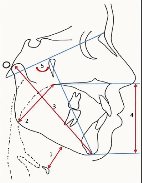
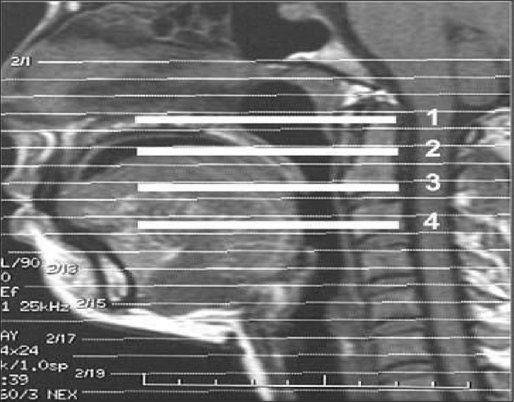
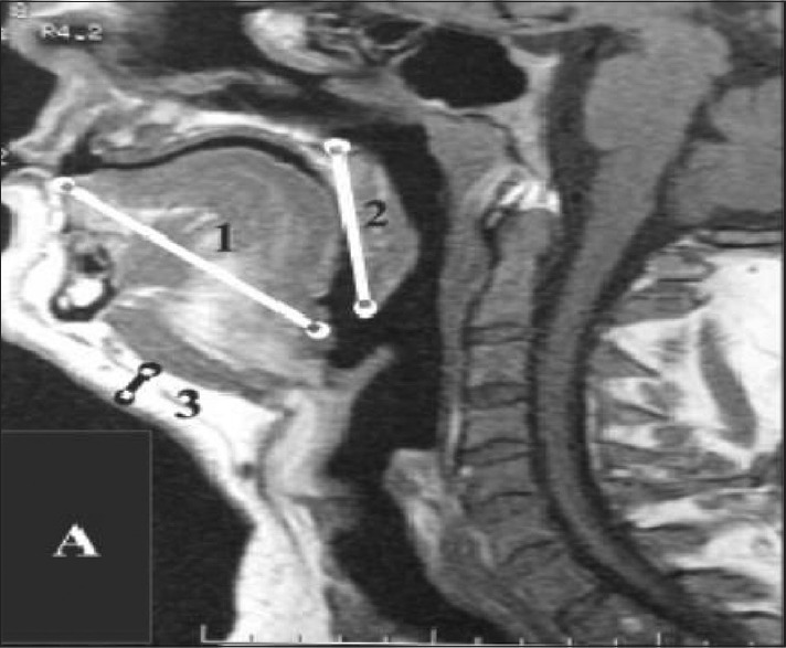
Forty cases and an equal number of matched controls were recruited. On X-ray cephalometry, it was observed that the cases had a significantly larger hyoid mandibular distance and soft palate length; and shorter mandibular length. The MRI cephalometric variables were significantly different, the soft palate length, tongue length, and submental fat were longer while the retropalatal and retroglossal distance was shorter amongst the cases. A statistically significant positive correlation was found between the cephalometric parameters and the indices of severity of OSA. An increased hyoid mandibular distance and soft palate length, and a decrease in the lower anterior facial height were found to be predictive of severe OSA (Apnea-Hypopnea Index ->30/h). An increased hyoid mandibular distance, soft palate length, and the tongue length and a reduced mandibular length were found to be predictive of need for continuous positive airway pressure (CPAP) pressures of ≥15 cm HO. There were significant differences between the cephalometric parameters of the Indian OSA patients and patients from other ethnicities reported in the literature.
OSA patients had a significantly smaller upper airway compared to age-, sex-, and body mass index-matched controls and cephalometric variables correlated with the indices of OSA severity. The cephalometric assessment was also predictive of severe OSA and the need for higher pressures of CPAP. This indicates the important role of upper airway anatomy in the pathogenesis of OSA.
上气道成像通常能够识别睡眠呼吸暂停的解剖学危险因素,并为阻塞性睡眠呼吸暂停(OSA)的病理生理学提供充分的见解。
我们在印度北部的一家三级护理医院进行了一项病例对照观察研究。所有病例和对照均接受了头颅侧位测量和磁共振成像(MRI),以评估颅面和上气道情况。只有病例进行了多导睡眠图测试,以确诊OSA并评估疾病严重程度。
招募了40例病例和数量相等的匹配对照。在X线头颅侧位测量中,观察到病例的舌骨下颌距离和软腭长度明显更大,而下颌长度更短。MRI头颅测量变量存在显著差异,病例组的软腭长度、舌长度和颏下脂肪更长,而后腭后和舌后距离更短。头颅测量参数与OSA严重程度指数之间存在统计学上显著的正相关。舌骨下颌距离和软腭长度增加,以及下前面部高度降低被发现可预测重度OSA(呼吸暂停低通气指数->30次/小时)。舌骨下颌距离、软腭长度和舌长度增加以及下颌长度减少被发现可预测需要持续气道正压通气(CPAP)压力≥15 cm H₂O。印度OSA患者的头颅测量参数与文献中报道的其他种族患者存在显著差异。
与年龄、性别和体重指数匹配的对照相比,OSA患者的上气道明显更小,且头颅测量变量与OSA严重程度指数相关。头颅测量评估也可预测重度OSA以及对更高CPAP压力的需求。这表明上气道解剖结构在OSA发病机制中具有重要作用。