• 文献检索
  • 文档翻译
  • 深度研究
  • 学术资讯
  • Suppr Zotero 插件Zotero 插件
  • 邀请有礼
  • 套餐&价格
  • 历史记录
应用&插件
Suppr Zotero 插件Zotero 插件浏览器插件Mac 客户端Windows 客户端微信小程序
定价
高级版会员购买积分包购买API积分包
服务
文献检索文档翻译深度研究API 文档MCP 服务
关于我们
关于 Suppr公司介绍联系我们用户协议隐私条款
关注我们

Suppr 超能文献

核心技术专利:CN118964589B侵权必究
粤ICP备2023148730 号-1Suppr @ 2026

文献检索

告别复杂PubMed语法,用中文像聊天一样搜索,搜遍4000万医学文献。AI智能推荐,让科研检索更轻松。

立即免费搜索

文件翻译

保留排版,准确专业,支持PDF/Word/PPT等文件格式,支持 12+语言互译。

免费翻译文档

深度研究

AI帮你快速写综述,25分钟生成高质量综述,智能提取关键信息,辅助科研写作。

立即免费体验

蓝光自发荧光眼底成像用于监测皇家外科学院大鼠的视网膜变性

Blue Autofluorescence Fundus Imaging for Monitoring Retinal Degeneration in Royal College of Surgeons Rats.

作者信息

Bubis Ettel, Sher Ifat, Skaat Alon, Sharvit-Ginon Inbal, Szalapak Alicja M, Moroz Iris, Kalter-Leibovici Ofra, Rotenstreich Ygal

机构信息

Goldschleger Eye Institute, Sheba Medical Center, Tel-Hashomer, Israel.

Sackler Faculty of Medicine, Tel Aviv University, Tel Aviv, Israel.

出版信息

Transl Vis Sci Technol. 2019 Feb 28;8(1):26. doi: 10.1167/tvst.8.1.26. eCollection 2019 Jan.

DOI:10.1167/tvst.8.1.26
PMID:30834174
原文链接:https://pmc.ncbi.nlm.nih.gov/articles/PMC6396687/
Abstract

PURPOSE

Development of a method for noninvasive longitudinal follow-up of retinal degeneration in the whole retina for Royal College of Surgeons (RCS) rats, a commonly used model of retinitis pigmentosa associated with mutations in the MER-proto-oncogene tyrosine kinase () gene.

METHODS

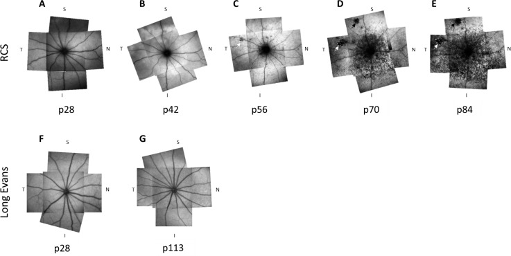
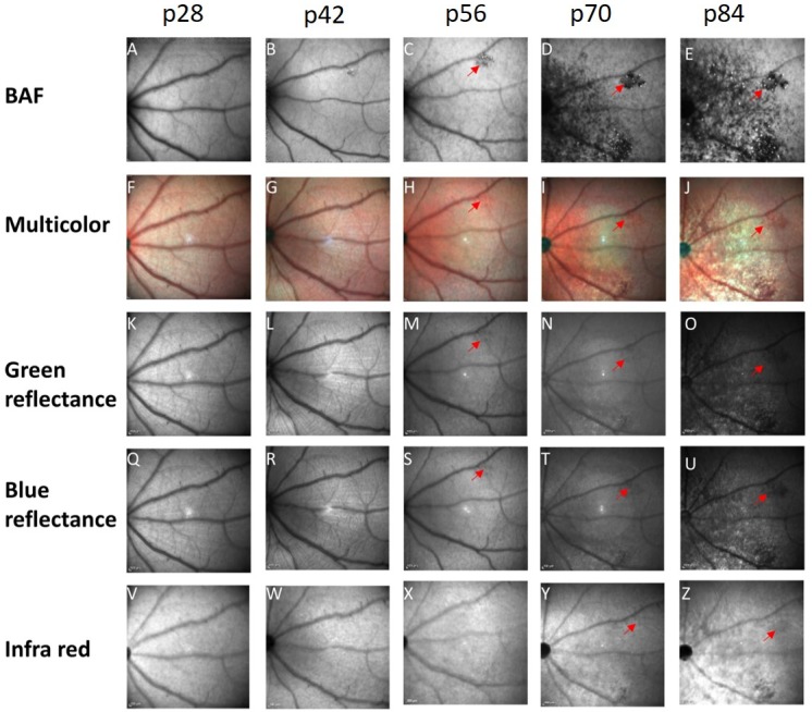
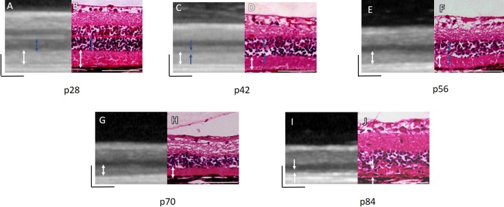
Pigmented RCS rats at postnatal (p) days p28 to p84 were subjected to a biweekly spectral-domain optical coherence tomography (SD-OCT), blue laser fundus autofluorescence (BL-FAF) imaging, and multicolor fundus imaging. Wild-type (WT; Long Evans) rats were tested as control.

RESULTS

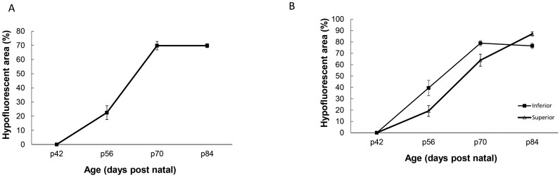
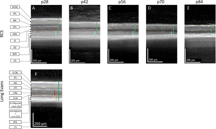
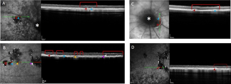
Hyperautofluorescence developed throughout the fundus at p42, concomitant with a significant increase in SD-OCT thickness and reflectivity of the debris zone (DZ) layer as well as thinning of the photoreceptor outer nuclear layer (ONL). From p56 to p84, discrete hypofluorescent lesions surrounded by hyperfluorescent flecks were demonstrated around the optic disc that gradually spread throughout the retina. The hypofluorescent lesions were associated with loss of ONL and gradual thinning of the DZ layer. No hypofluorescent BL-FAF lesions were observed in WT rats.

CONCLUSIONS

This study suggests that BL-FAF imaging may present a new method for noninvasive longitudinal follow-up of retinal degeneration in nearly the whole retina in RCS rats.

TRANSLATIONAL RELEVANCE

A clinical test was developed that may be implemented in translational studies in the RCS rat model of -associated retinitis pigmentosa.

摘要

目的

开发一种用于对皇家外科学院(RCS)大鼠整个视网膜的视网膜变性进行无创纵向随访的方法,RCS大鼠是一种常用的与MER原癌基因酪氨酸激酶()基因突变相关的色素性视网膜炎模型。

方法

对出生后(p)28至84天的色素性RCS大鼠每两周进行一次光谱域光学相干断层扫描(SD-OCT)、蓝色激光眼底自发荧光(BL-FAF)成像和多色眼底成像。以野生型(WT;长Evans)大鼠作为对照进行检测。

结果

在p42时整个眼底出现高自发荧光,同时SD-OCT显示碎片带(DZ)层厚度和反射率显著增加,以及光感受器外核层(ONL)变薄。从p56到p84,视盘周围出现由高荧光斑点包围的离散低荧光病变,并逐渐扩散至整个视网膜。这些低荧光病变与ONL的丧失和DZ层的逐渐变薄有关。在WT大鼠中未观察到低荧光的BL-FAF病变。

结论

本研究表明,BL-FAF成像可能为RCS大鼠几乎整个视网膜的视网膜变性无创纵向随访提供一种新方法。

转化相关性

开发了一种临床试验,可在与相关色素性视网膜炎的RCS大鼠模型的转化研究中实施。

https://cdn.ncbi.nlm.nih.gov/pmc/blobs/f2ac/6396687/30fac905e24f/i2164-2591-8-1-26-f06.jpg
https://cdn.ncbi.nlm.nih.gov/pmc/blobs/f2ac/6396687/a5f7bec1d048/i2164-2591-8-1-26-f01.jpg
https://cdn.ncbi.nlm.nih.gov/pmc/blobs/f2ac/6396687/c1c0135937e5/i2164-2591-8-1-26-f02.jpg
https://cdn.ncbi.nlm.nih.gov/pmc/blobs/f2ac/6396687/9807c57aa407/i2164-2591-8-1-26-f03.jpg
https://cdn.ncbi.nlm.nih.gov/pmc/blobs/f2ac/6396687/a720b7b66687/i2164-2591-8-1-26-f04.jpg
https://cdn.ncbi.nlm.nih.gov/pmc/blobs/f2ac/6396687/fb58d9b49697/i2164-2591-8-1-26-f05.jpg
https://cdn.ncbi.nlm.nih.gov/pmc/blobs/f2ac/6396687/30fac905e24f/i2164-2591-8-1-26-f06.jpg

相似文献

1
Blue Autofluorescence Fundus Imaging for Monitoring Retinal Degeneration in Royal College of Surgeons Rats.蓝光自发荧光眼底成像用于监测皇家外科学院大鼠的视网膜变性
Transl Vis Sci Technol. 2019 Feb 28;8(1):26. doi: 10.1167/tvst.8.1.26. eCollection 2019 Jan.
2
Long-term Characterization of Retinal Degeneration in Royal College of Surgeons Rats Using Spectral-Domain Optical Coherence Tomography.使用频域光学相干断层扫描技术对皇家外科医学院大鼠视网膜变性进行长期特征分析。
Invest Ophthalmol Vis Sci. 2017 Mar 1;58(3):1378-1386. doi: 10.1167/iovs.16-20363.
3
Evaluation of Retinal Function and Morphology of the Pink-Eyed Royal College of Surgeons (RCS) Rat: A Comparative Study of in Vivo and in Vitro Methods.粉红眼皇家外科学院(RCS)大鼠视网膜功能与形态学评估:体内与体外方法的比较研究
Curr Eye Res. 2017 Feb;42(2):273-281. doi: 10.1080/02713683.2016.1179333. Epub 2016 Jun 30.
4
Quantification of microvascular change of retinal degeneration in Royal College of Surgeons rats using high-resolution spectral domain optical coherence tomography angiography.利用高分辨率谱域光相干断层扫描血管造影术定量评估皇家外科学院大鼠视网膜变性的微血管变化。
J Biomed Opt. 2023 Oct;28(10):106001. doi: 10.1117/1.JBO.28.10.106001. Epub 2023 Oct 13.
5
Dorsal-Ventral Differences in Retinal Structure in the Pigmented Royal College of Surgeons Model of Retinal Degeneration.色素性皇家外科学院视网膜变性模型中视网膜结构的背腹差异
Front Cell Neurosci. 2021 Jan 18;14:553708. doi: 10.3389/fncel.2020.553708. eCollection 2020.
6
Long time remodeling during retinal degeneration evaluated by optical coherence tomography, immunocytochemistry and fundus autofluorescence.通过光学相干断层扫描、免疫细胞化学和眼底自发荧光评估视网膜变性过程中的长期重塑。
Exp Eye Res. 2016 Sep;150:122-34. doi: 10.1016/j.exer.2015.10.012. Epub 2015 Oct 29.
7
Microglia Inhibition Delays Retinal Degeneration Due to MerTK Phagocytosis Receptor Deficiency.小胶质细胞抑制因 MerTK 吞噬受体缺陷导致的视网膜变性。
Front Immunol. 2020 Jul 16;11:1463. doi: 10.3389/fimmu.2020.01463. eCollection 2020.
8
Optical Coherence Tomography of Retinal Degeneration in Royal College of Surgeons Rats and Its Correlation with Morphology and Electroretinography.皇家外科学院大鼠视网膜变性的光学相干断层扫描及其与形态学和视网膜电图的相关性
PLoS One. 2016 Sep 19;11(9):e0162835. doi: 10.1371/journal.pone.0162835. eCollection 2016.
9
Longitudinal Structural and Microvascular Observation in RCS Rat Eyes Using Optical Coherence Tomography Angiography.应用光相干断层扫描血管造影术观察 RCS 大鼠眼部的纵向结构和微血管。
Invest Ophthalmol Vis Sci. 2020 Jun 3;61(6):54. doi: 10.1167/iovs.61.6.54.
10
In-vivo mapping of drusen by fundus autofluorescence and spectral-domain optical coherence tomography imaging.通过眼底自发荧光和光谱域光学相干断层扫描成像对玻璃膜疣进行体内成像。
Graefes Arch Clin Exp Ophthalmol. 2016 Jan;254(1):59-67. doi: 10.1007/s00417-015-3012-4. Epub 2015 Apr 24.

引用本文的文献

1
Housing RCS rats under specific pathogen-free conditions mildly ameliorates retinal degeneration and alters intestine microbiota.在特定病原体-free条件下饲养RCS大鼠可轻度改善视网膜变性并改变肠道微生物群。
Sci Rep. 2024 Sep 16;14(1):21583. doi: 10.1038/s41598-024-70960-z.
2
Unified modeling of photothermal and photochemical damage.光热损伤与光化学损伤的统一建模
Front Ophthalmol (Lausanne). 2024 Aug 19;4:1408869. doi: 10.3389/fopht.2024.1408869. eCollection 2024.
3
An extraocular electrical stimulation approach to slow down the progression of retinal degeneration in an animal model.

本文引用的文献

1
Long-term Rescue of Photoreceptors in a Rodent Model of Retinitis Pigmentosa Associated with MERTK Mutation.与 MERTK 突变相关的视网膜色素变性的啮齿动物模型中感光细胞的长期挽救。
Sci Rep. 2018 Jul 27;8(1):11312. doi: 10.1038/s41598-018-29631-z.
2
Otx2-Genetically Modified Retinal Pigment Epithelial Cells Rescue Photoreceptors after Transplantation.Otx2 基因修饰的视网膜色素上皮细胞移植后可挽救光感受器。
Mol Ther. 2018 Jan 3;26(1):219-237. doi: 10.1016/j.ymthe.2017.09.007. Epub 2017 Sep 8.
3
Optical coherence tomography: fundamental principles, instrumental designs and biomedical applications.
一种眼外电刺激方法,可减缓动物模型中视网膜变性的进展。
Sci Rep. 2023 Sep 23;13(1):15924. doi: 10.1038/s41598-023-40547-1.
4
retinal imaging in translational regenerative research.转化性再生研究中的视网膜成像
Ann Transl Med. 2020 Sep;8(17):1096. doi: 10.21037/atm-20-4355.
5
Systemic Treatment With Nicotinamide Riboside Is Protective in a Mouse Model of Light-Induced Retinal Degeneration.烟酰胺核糖苷全身治疗对光诱导的视网膜退行性变小鼠模型具有保护作用。
Invest Ophthalmol Vis Sci. 2020 Aug 3;61(10):47. doi: 10.1167/iovs.61.10.47.
6
Wheel running exercise protects against retinal degeneration in the I307N rhodopsin mouse model of inducible autosomal dominant retinitis pigmentosa.轮转运动可预防可诱导的常染色体显性遗传性视网膜色素变性的I307N视紫红质小鼠模型中的视网膜变性。
Mol Vis. 2019 Aug 21;25:462-476. eCollection 2019.
光学相干断层扫描:基本原理、仪器设计及生物医学应用
Biophys Rev. 2011 Sep;3(3):155. doi: 10.1007/s12551-011-0054-7. Epub 2011 Aug 6.
4
Long-term Characterization of Retinal Degeneration in Royal College of Surgeons Rats Using Spectral-Domain Optical Coherence Tomography.使用频域光学相干断层扫描技术对皇家外科医学院大鼠视网膜变性进行长期特征分析。
Invest Ophthalmol Vis Sci. 2017 Mar 1;58(3):1378-1386. doi: 10.1167/iovs.16-20363.
5
Clinical applications of fundus autofluorescence in retinal disease.眼底自发荧光在视网膜疾病中的临床应用
Int J Retina Vitreous. 2016 Apr 8;2:12. doi: 10.1186/s40942-016-0035-x. eCollection 2016.
6
Fundus Autofluorescence in Age-related Macular Degeneration.年龄相关性黄斑变性中的眼底自发荧光
Optom Vis Sci. 2017 Feb;94(2):246-259. doi: 10.1097/OPX.0000000000000997.
7
Optical Coherence Tomography of Retinal Degeneration in Royal College of Surgeons Rats and Its Correlation with Morphology and Electroretinography.皇家外科学院大鼠视网膜变性的光学相干断层扫描及其与形态学和视网膜电图的相关性
PLoS One. 2016 Sep 19;11(9):e0162835. doi: 10.1371/journal.pone.0162835. eCollection 2016.
8
Treatment of retinitis pigmentosa due to MERTK mutations by ocular subretinal injection of adeno-associated virus gene vector: results of a phase I trial.通过眼内视网膜下注射腺相关病毒基因载体治疗因MERTK突变引起的色素性视网膜炎:I期试验结果
Hum Genet. 2016 Mar;135(3):327-43. doi: 10.1007/s00439-016-1637-y. Epub 2016 Jan 29.
9
Expanding the clinical, allelic, and locus heterogeneity of retinal dystrophies.扩大视网膜营养不良的临床、等位基因和基因座异质性。
Genet Med. 2016 Jun;18(6):554-62. doi: 10.1038/gim.2015.127. Epub 2015 Sep 10.
10
Fundus Autofluorescence and RPE Lipofuscin in Age-Related Macular Degeneration.年龄相关性黄斑变性中的眼底自发荧光与视网膜色素上皮脂褐质
J Clin Med. 2014;3(4):1302-21. doi: 10.3390/jcm3041302.