Jin He, He Miao, Liu Hongshan, Zhong Xiaoying, Wu Junshu, Liu Liangping, Ding Hui, Zhang Chi, Zhong Xingwu
State Key Laboratory of Ophthalmology, Zhongshan Ophthalmic Center, Sun Yat-sen University, Guangzhou, China.
Affiliated Hospital of Guilin Medical University, Guilin Medical University, Guilin, China.
Cornea. 2019 Apr;38(4):446-453. doi: 10.1097/ICO.0000000000001877.
To evaluate the feasibility and efficacy of small-incision femtosecond laser-assisted intracorneal concave lenticule implantation (SFII) and penetrating keratoplasty (PKP) in patients with progressive keratoconus.
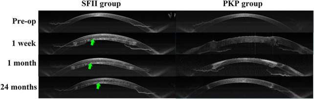
All the patients were clinically diagnosed with progressive keratoconus. Twenty patients underwent PKP (PKP group), and 11 patients underwent SFII (SFII group). Visual acuity, intraocular pressure, corneal topography, corneal visualization Scheimpflug technology, anterior segment optical coherence tomography, and in vivo confocal microscopy were analyzed.
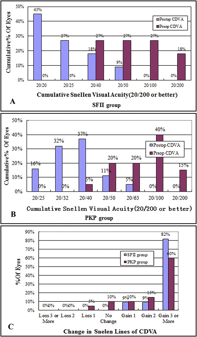
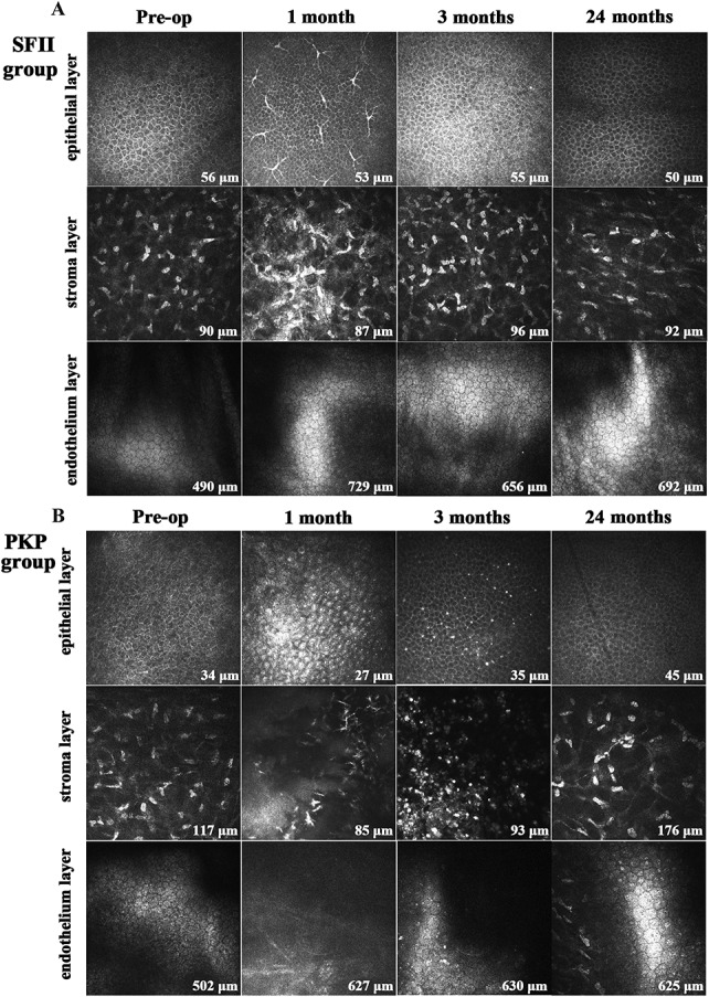
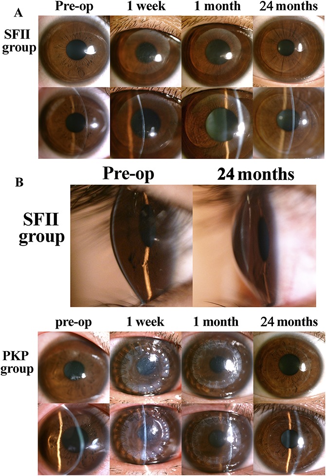
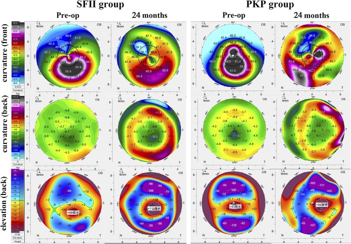
Vision improved at 3 months postoperatively in the SFII group. In the PKP group, corrected distance visual acuity improved 1 week after surgery. Corneal topography showed a statistically significant decrease in the anterior K1 and K2. Corneal visualization Scheimpflug technology showed that changes in the biomechanical parameters of the SFII group were also statistically different from those of the PKP group. All the grafts from both groups were clearly visible by anterior segment optical coherence tomography observation. The central corneal thickness of both groups was stable during the 24-month study period. In vivo confocal microscopy showed a few dendritic cells in the subepithelial region in the SFII group. At 3 months after surgery, many dendritic cells and inflammatory cells were observed in the basal epithelium and stroma in the PKP group.
Both SFII and PKP surgical procedures resulted in a stable corneal volume and improved visual acuity in this long-term study. SFII was less invasive and more efficient compared with PKP.
评估小切口飞秒激光辅助角膜基质内凹透镜植入术(SFII)和穿透性角膜移植术(PKP)治疗进行性圆锥角膜患者的可行性和疗效。
所有患者均经临床诊断为进行性圆锥角膜。20例患者接受PKP(PKP组),11例患者接受SFII(SFII组)。分析视力、眼压、角膜地形图、角膜可视化Scheimpflug技术、眼前节光学相干断层扫描和活体共聚焦显微镜检查结果。
SFII组术后3个月视力提高。PKP组术后1周矫正远视力提高。角膜地形图显示前表面K1和K2有统计学意义的降低。角膜可视化Scheimpflug技术显示SFII组生物力学参数的变化与PKP组也有统计学差异。两组所有移植片经眼前节光学相干断层扫描观察均清晰可见。在24个月的研究期间,两组中央角膜厚度均稳定。活体共聚焦显微镜检查显示SFII组上皮下区域有少量树突状细胞。术后3个月,PKP组基底上皮和基质中观察到许多树突状细胞和炎性细胞。
在这项长期研究中,SFII和PKP手术均能使角膜容积稳定并提高视力。与PKP相比,SFII侵入性更小且更有效。