• 文献检索
  • 文档翻译
  • 深度研究
  • 学术资讯
  • Suppr Zotero 插件Zotero 插件
  • 邀请有礼
  • 套餐&价格
  • 历史记录
应用&插件
Suppr Zotero 插件Zotero 插件浏览器插件Mac 客户端Windows 客户端微信小程序
定价
高级版会员购买积分包购买API积分包
服务
文献检索文档翻译深度研究API 文档MCP 服务
关于我们
关于 Suppr公司介绍联系我们用户协议隐私条款
关注我们

Suppr 超能文献

核心技术专利:CN118964589B侵权必究
粤ICP备2023148730 号-1Suppr @ 2026

文献检索

告别复杂PubMed语法,用中文像聊天一样搜索,搜遍4000万医学文献。AI智能推荐,让科研检索更轻松。

立即免费搜索

文件翻译

保留排版,准确专业,支持PDF/Word/PPT等文件格式,支持 12+语言互译。

免费翻译文档

深度研究

AI帮你快速写综述,25分钟生成高质量综述,智能提取关键信息,辅助科研写作。

立即免费体验

社区环境下乳腺癌的磁化转移磁共振成像:新辅助治疗中的可重复性及初步结果

Magnetization Transfer MRI of Breast Cancer in the Community Setting: Reproducibility and Preliminary Results in Neoadjuvant Therapy.

作者信息

Virostko John, Sorace Anna G, Wu Chengyue, Ekrut David, Jarrett Angela M, Upadhyaya Raghave M, Avery Sarah, Patt Debra, Goodgame Boone, Yankeelov Thomas E

机构信息

Department of Diagnostic Medicine.

Livestrong Cancer Institutes.

出版信息

Tomography. 2019 Mar;5(1):44-52. doi: 10.18383/j.tom.2018.00019.

DOI:10.18383/j.tom.2018.00019
PMID:30854441
原文链接:https://pmc.ncbi.nlm.nih.gov/articles/PMC6403021/
Abstract

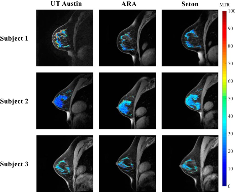
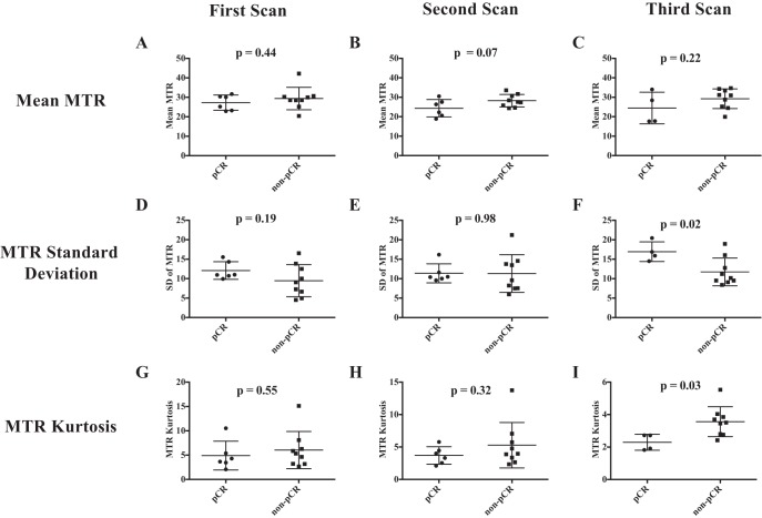
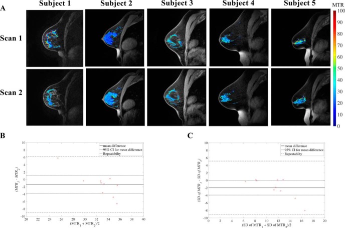
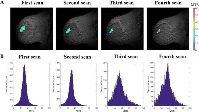
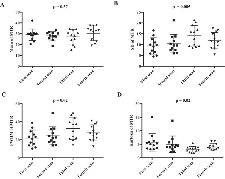
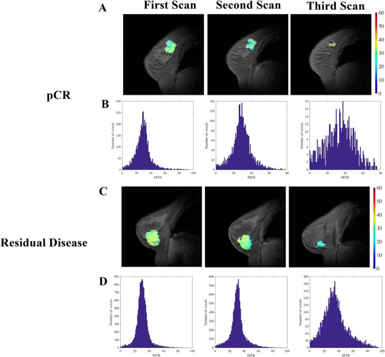
Repeatability and reproducibility of magnetization transfer magnetic resonance imaging of the breast, and the ability of this technique to assess the response of locally advanced breast cancer to neoadjuvant therapy (NAT), are determined. Reproducibility scans at 3 different 3 T scanners, including 2 scanners in community imaging centers, found a 16.3% difference (n = 3) in magnetization transfer ratio (MTR) in healthy breast fibroglandular tissue. Repeatability scans (n = 10) found a difference of ∼8.1% in the MTR measurement of fibroglandular tissue between the 2 measurements. Thus, MTR is repeatable and reproducible in the breast and can be integrated into community imaging clinics. Serial magnetization transfer magnetic resonance imaging performed at longitudinal time points during NAT indicated no significant change in average tumoral MTR during treatment. However, histogram analysis indicated an increase in the dispersion of MTR values of the tumor during NAT, as quantified by higher standard deviation ( = .005), higher full width at half maximum ( = .02), and lower kurtosis ( = .02). Patients' stratification into those with pathological complete response (pCR; n = 6) at the conclusion of NAT and those with residual disease (n = 9) showed wider distribution of tumor MTR values in patients who achieved pCR after 2-4 cycles of NAT, as quantified by higher standard deviation ( = .02), higher full width at half maximum ( = .03), and lower kurtosis ( = .03). Thus, MTR can be used as an imaging metric to assess response to breast NAT.

摘要

确定了乳腺磁化传递磁共振成像的可重复性和再现性,以及该技术评估局部晚期乳腺癌对新辅助治疗(NAT)反应的能力。在3台不同的3T扫描仪上进行的再现性扫描,包括社区影像中心的2台扫描仪,发现健康乳腺纤维腺组织的磁化传递率(MTR)存在16.3%的差异(n = 3)。重复性扫描(n = 10)发现两次测量之间纤维腺组织的MTR测量值相差约8.1%。因此,MTR在乳腺中具有可重复性和再现性,可整合到社区影像诊所中。在NAT期间的纵向时间点进行的系列磁化传递磁共振成像表明,治疗期间肿瘤平均MTR无显著变化。然而,直方图分析表明,NAT期间肿瘤MTR值的离散度增加,通过更高的标准差( = 0.005)、更高的半高宽( = 0.02)和更低的峰度( = 0.02)来量化。将患者分层为NAT结束时达到病理完全缓解(pCR;n = 6)的患者和有残留疾病的患者(n = 9),结果显示,在接受2 - 4个周期NAT后达到pCR的患者中,肿瘤MTR值分布更宽,通过更高的标准差( = 0.02)、更高的半高宽( = 0.03)和更低的峰度( = 0.03)来量化。因此,MTR可作为一种成像指标来评估乳腺NAT的反应。

https://cdn.ncbi.nlm.nih.gov/pmc/blobs/47b0/6403021/60d12c8f2950/tom0041801250006.jpg
https://cdn.ncbi.nlm.nih.gov/pmc/blobs/47b0/6403021/baaabbb16f99/tom0041801250001.jpg
https://cdn.ncbi.nlm.nih.gov/pmc/blobs/47b0/6403021/f8f7cb1d2934/tom0041801250002.jpg
https://cdn.ncbi.nlm.nih.gov/pmc/blobs/47b0/6403021/c590136e7aa7/tom0041801250003.jpg
https://cdn.ncbi.nlm.nih.gov/pmc/blobs/47b0/6403021/a4bed2e56201/tom0041801250004.jpg
https://cdn.ncbi.nlm.nih.gov/pmc/blobs/47b0/6403021/4300658aa13d/tom0041801250005.jpg
https://cdn.ncbi.nlm.nih.gov/pmc/blobs/47b0/6403021/60d12c8f2950/tom0041801250006.jpg

相似文献

1
Magnetization Transfer MRI of Breast Cancer in the Community Setting: Reproducibility and Preliminary Results in Neoadjuvant Therapy.社区环境下乳腺癌的磁化转移磁共振成像:新辅助治疗中的可重复性及初步结果
Tomography. 2019 Mar;5(1):44-52. doi: 10.18383/j.tom.2018.00019.
2
Comparison of automated breast volume scanning with conventional ultrasonography, mammography, and MRI to assess residual breast cancer after neoadjuvant therapy by molecular type.通过分子类型比较自动乳腺容积扫描与传统超声、乳腺X线摄影及磁共振成像在评估新辅助治疗后残余乳腺癌方面的差异。
Clin Radiol. 2023 May;78(5):e393-e400. doi: 10.1016/j.crad.2022.12.002. Epub 2023 Jan 13.
3
Magnetization transfer ratio: a potential biomarker for the assessment of postradiation fibrosis in patients with rectal cancer.磁化传递率:评估直肠癌放疗后纤维化的潜在生物标志物。
Invest Radiol. 2014 Jan;49(1):29-34. doi: 10.1097/RLI.0b013e3182a3459b.
4
The rate of breast fibroglandular enhancement during dynamic contrast-enhanced MRI reflects response to neoadjuvant therapy.动态对比增强 MRI 中乳腺纤维腺体增强率反映了对新辅助治疗的反应。
Eur J Radiol. 2021 Mar;136:109534. doi: 10.1016/j.ejrad.2021.109534. Epub 2021 Jan 9.
5
Three-dimensional ultrasonography of the breast; An adequate replacement for MRI in neoadjuvant chemotherapy tumour response evaluation? - RESPONDER trial.乳腺三维超声;新辅助化疗肿瘤反应评估中 MRI 的充分替代品? - RESPONDER 试验。
Eur J Radiol. 2018 Jul;104:94-100. doi: 10.1016/j.ejrad.2018.05.005. Epub 2018 May 5.
6
Histogram analysis of quantitative T1 and MT maps from ultrahigh field MRI in clinically isolated syndrome and relapsing-remitting multiple sclerosis.超高场磁共振成像定量T1和MT图在临床孤立综合征和复发缓解型多发性硬化中的直方图分析
NMR Biomed. 2015 Nov;28(11):1374-82. doi: 10.1002/nbm.3385. Epub 2015 Sep 7.
7
Radiologic complete response (rCR) in contrast-enhanced magnetic resonance imaging (CE-MRI) after neoadjuvant chemotherapy for early breast cancer predicts recurrence-free survival but not pathologic complete response (pCR).新辅助化疗后对比增强磁共振成像(CE-MRI)的放射学完全缓解(rCR)可预测无复发生存期,但不能预测病理完全缓解(pCR)。
Breast Cancer Res. 2019 Jan 31;21(1):19. doi: 10.1186/s13058-018-1091-y.
8
Systematic variation of off-resonance prepulses for clinical magnetization transfer contrast imaging at 0.2, 1.5, and 3.0 tesla.用于0.2、1.5和3.0特斯拉临床磁共振波谱成像的失谐预脉冲的系统变化
Invest Radiol. 2008 Jan;43(1):16-26. doi: 10.1097/RLI.0b013e3181559949.
9
Magnetization transfer imaging to assess tumour response after chemoradiotherapy in rectal cancer.磁共振磁化传递成像评估直肠癌放化疗后的肿瘤反应
Eur Radiol. 2016 Feb;26(2):390-7. doi: 10.1007/s00330-015-3856-3. Epub 2015 Jun 12.
10
Magnetic resonance imaging evaluation of residual tumors in breast cancer after neoadjuvant chemotherapy: surgical implications.新辅助化疗后乳腺癌残留肿瘤的磁共振成像评估:手术意义
Acta Radiol. 2016 May;57(5):529-37. doi: 10.1177/0284185115597263. Epub 2015 Jul 31.

引用本文的文献

1
Quantitative magnetic resonance imaging and tumor forecasting of breast cancer patients in the community setting.社区环境下乳腺癌患者的定量磁共振成像与肿瘤预测。
Nat Protoc. 2021 Nov;16(11):5309-5338. doi: 10.1038/s41596-021-00617-y. Epub 2021 Sep 22.
2
Quantitative Imaging Enters the Clinical Arena: A Personal Viewpoint.定量成像进入临床领域:个人观点。
Tomography. 2020 Jun;6(2):56-59. doi: 10.18383/j.tom.2020.00028.
3
Current and Emerging Magnetic Resonance-Based Techniques for Breast Cancer.用于乳腺癌的当前及新兴的基于磁共振成像的技术

本文引用的文献

1
Repeatability, reproducibility, and accuracy of quantitative mri of the breast in the community radiology setting.社区放射学环境下乳腺定量MRI的可重复性、再现性及准确性
J Magn Reson Imaging. 2018 Mar 23. doi: 10.1002/jmri.26011.
2
Modelling and interpretation of magnetization transfer imaging in the brain.脑磁化转移成像的建模与解释。
Neuroimage. 2018 Nov 15;182:128-135. doi: 10.1016/j.neuroimage.2017.11.065. Epub 2017 Dec 5.
3
Dynamic contrast-enhanced magnetic resonance imaging and diffusion-weighted magnetic resonance imaging for predicting the response of locally advanced breast cancer to neoadjuvant therapy: a meta-analysis.
Front Med (Lausanne). 2020 May 12;7:175. doi: 10.3389/fmed.2020.00175. eCollection 2020.
4
Bimodal magnetic resonance and optical imaging of extracellular matrix remodelling by orthotopic ovarian tumours.通过原位卵巢肿瘤的磁共振和光学双模成像观察细胞外基质重塑。
Br J Cancer. 2020 Jul;123(2):216-225. doi: 10.1038/s41416-020-0878-7. Epub 2020 May 11.
动态对比增强磁共振成像和扩散加权磁共振成像预测局部晚期乳腺癌对新辅助治疗反应的Meta分析
J Med Imaging (Bellingham). 2018 Jan;5(1):011011. doi: 10.1117/1.JMI.5.1.011011. Epub 2017 Nov 24.
4
Quantitative Magnetization Transfer Imaging of the Breast at 3.0 T: Reproducibility in Healthy Volunteers.3.0T乳腺定量磁化转移成像:健康志愿者中的可重复性
Tomography. 2016 Dec;2(4):260-266. doi: 10.18383/j.tom.2016.00142.
5
From transformation to metastasis: deconstructing the extracellular matrix in breast cancer.从转化到转移:解析乳腺癌中的细胞外基质
Cancer Metastasis Rev. 2016 Dec;35(4):655-667. doi: 10.1007/s10555-016-9650-0.
6
Histogram analysis of quantitative T1 and MT maps from ultrahigh field MRI in clinically isolated syndrome and relapsing-remitting multiple sclerosis.超高场磁共振成像定量T1和MT图在临床孤立综合征和复发缓解型多发性硬化中的直方图分析
NMR Biomed. 2015 Nov;28(11):1374-82. doi: 10.1002/nbm.3385. Epub 2015 Sep 7.
7
Magnetization transfer imaging to assess tumour response after chemoradiotherapy in rectal cancer.磁共振磁化传递成像评估直肠癌放化疗后的肿瘤反应
Eur Radiol. 2016 Feb;26(2):390-7. doi: 10.1007/s00330-015-3856-3. Epub 2015 Jun 12.
8
Chemotherapy induces the cancer-associated fibroblast phenotype, activating paracrine Hedgehog-GLI signalling in breast cancer cells.化疗诱导癌症相关成纤维细胞表型,激活乳腺癌细胞中的旁分泌刺猬索尼克-胶质瘤相关蛋白信号通路。
Oncotarget. 2015 May 10;6(13):10728-45. doi: 10.18632/oncotarget.3828.
9
Evaluation of the treatment response to neoadjuvant chemotherapy in locally advanced breast cancer using combined magnetic resonance vascular maps and apparent diffusion coefficient.使用联合磁共振血管图和表观扩散系数评估局部晚期乳腺癌新辅助化疗的治疗反应
J Magn Reson Imaging. 2015 Nov;42(5):1407-20. doi: 10.1002/jmri.24915. Epub 2015 Apr 15.
10
Multiparametric magnetic resonance imaging for predicting pathological response after the first cycle of neoadjuvant chemotherapy in breast cancer.多参数磁共振成像预测乳腺癌新辅助化疗第一周期后的病理反应
Invest Radiol. 2015 Apr;50(4):195-204. doi: 10.1097/RLI.0000000000000100.