Zhu Xiu-Liang, Su Wei-Wei, Tang Jin-Long, Yao Li-Ding, Lu Liang-Ji, Sun Xiu-Juan
Department of Radiology.
Department of Ultrasonography.
Medicine (Baltimore). 2019 Mar;98(10):e13416. doi: 10.1097/MD.0000000000013416.
Rhabdomyosarcoma (RMS) has known as a highly malignant soft tissue sarcoma, representing 5% to 10% of all solid tumors in childhood. Alveolar rhabdomyosarcoma (ARMS) of the retrorectal-presacral space is an extremely rare lesion for adult, no study has been reported in the English literature.
A 51-year-old male presented with abdominal pain for 1 month, significantly worse when having a bowel movement.
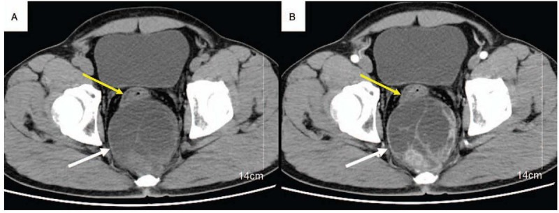
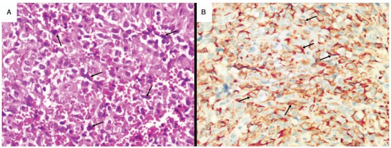
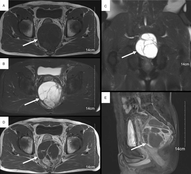
Computed tomography (CT) and magnetic resonance imaging (MRI) of the pelvis showed a solid-cystic, enhancing lesion of dimension located in retrorectal-presacral space. The surgical specimen was reported as ARMS after pathological evaluation.
The tumor was complete surgical resection, and after surgery, the patient was treated with combination chemotherapy.
At 23 months follow up, the patient was asymptomatic with no evidence of metastases or local recurrence.
Improvements in imaging in addition to early surgical intervention and chemotherapy treatment are crucial to improve survival chances against RMS.
横纹肌肉瘤(RMS)是一种高度恶性的软组织肉瘤,占儿童所有实体瘤的5%至10%。直肠后-骶前间隙的腺泡状横纹肌肉瘤(ARMS)在成人中极为罕见,英文文献中尚无相关研究报道。
一名51岁男性,腹痛1个月,排便时明显加重。
骨盆计算机断层扫描(CT)和磁共振成像(MRI)显示,直肠后-骶前间隙有一个实性-囊性、强化的肿块。病理评估后,手术标本报告为ARMS。
肿瘤完整切除,术后患者接受联合化疗。
随访23个月时,患者无症状,无转移或局部复发迹象。
除早期手术干预和化疗外,改善影像学检查对于提高抗RMS的生存几率至关重要。