• 文献检索
  • 文档翻译
  • 深度研究
  • 学术资讯
  • Suppr Zotero 插件Zotero 插件
  • 邀请有礼
  • 套餐&价格
  • 历史记录
应用&插件
Suppr Zotero 插件Zotero 插件浏览器插件Mac 客户端Windows 客户端微信小程序
定价
高级版会员购买积分包购买API积分包
服务
文献检索文档翻译深度研究API 文档MCP 服务
关于我们
关于 Suppr公司介绍联系我们用户协议隐私条款
关注我们

Suppr 超能文献

核心技术专利:CN118964589B侵权必究
粤ICP备2023148730 号-1Suppr @ 2026

文献检索

告别复杂PubMed语法,用中文像聊天一样搜索,搜遍4000万医学文献。AI智能推荐,让科研检索更轻松。

立即免费搜索

文件翻译

保留排版,准确专业,支持PDF/Word/PPT等文件格式,支持 12+语言互译。

免费翻译文档

深度研究

AI帮你快速写综述,25分钟生成高质量综述,智能提取关键信息,辅助科研写作。

立即免费体验

基于多图谱和深度学习方法的脑肿瘤患者危险器官自动勾画的能谱 CT 研究。

Dual-energy CT for automatic organs-at-risk segmentation in brain-tumor patients using a multi-atlas and deep-learning approach.

机构信息

Department of Radiation Oncology (MAASTRO), GROW-School for Oncology and Developmental Biology, Maastricht University Medical Centre, Maastricht, Netherlands.

OncoRay - National Center for Radiation Research in Oncology, Faculty of Medicine and University Hospital Carl Gustav Carus, Technische Universität Dresden, Helmholtz-Zentrum Dresden - Rossendorf, Dresden, Germany.

出版信息

Sci Rep. 2019 Mar 11;9(1):4126. doi: 10.1038/s41598-019-40584-9.

DOI:10.1038/s41598-019-40584-9
PMID:30858409
原文链接:https://pmc.ncbi.nlm.nih.gov/articles/PMC6411877/
Abstract

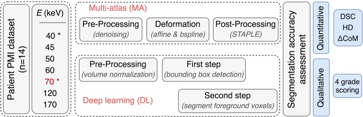
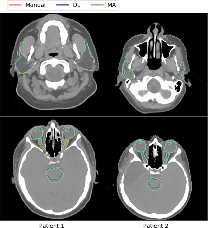
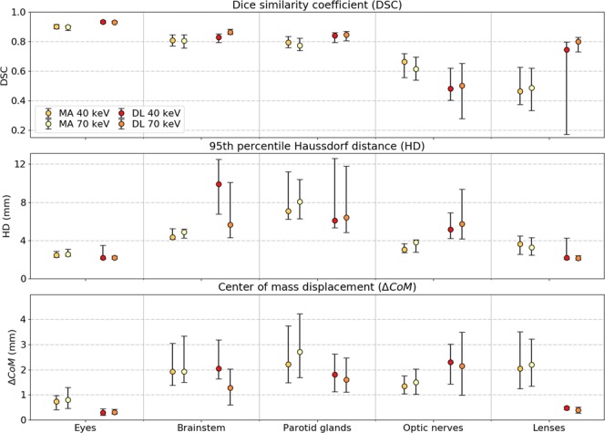
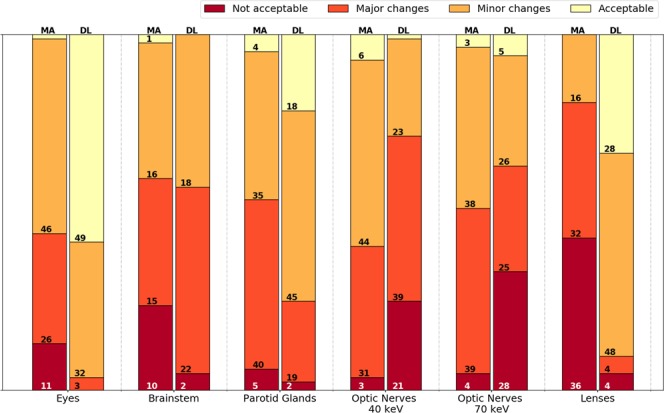
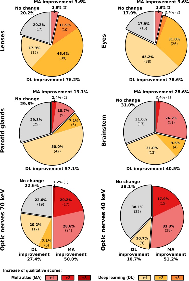
In radiotherapy, computed tomography (CT) datasets are mostly used for radiation treatment planning to achieve a high-conformal tumor coverage while optimally sparing healthy tissue surrounding the tumor, referred to as organs-at-risk (OARs). Based on CT scan and/or magnetic resonance images, OARs have to be manually delineated by clinicians, which is one of the most time-consuming tasks in the clinical workflow. Recent multi-atlas (MA) or deep-learning (DL) based methods aim to improve the clinical routine by an automatic segmentation of OARs on a CT dataset. However, so far no studies investigated the performance of these MA or DL methods on dual-energy CT (DECT) datasets, which have been shown to improve the image quality compared to conventional 120 kVp single-energy CT. In this study, the performance of an in-house developed MA and a DL method (two-step three-dimensional U-net) was quantitatively and qualitatively evaluated on various DECT-derived pseudo-monoenergetic CT datasets ranging from 40 keV to 170 keV. At lower energies, the MA method resulted in more accurate OAR segmentations. Both the qualitative and quantitative metric analysis showed that the DL approach often performed better than the MA method.

摘要

在放射治疗中,计算机断层扫描(CT)数据集大多用于放射治疗计划,以实现高适形肿瘤覆盖,同时最大限度地保护肿瘤周围的健康组织,即危及器官(OAR)。基于 CT 扫描和/或磁共振图像,临床医生必须手动勾画 OAR,这是临床工作流程中最耗时的任务之一。最近的多图谱(MA)或基于深度学习(DL)的方法旨在通过在 CT 数据集上自动分割 OAR 来改善临床常规。然而,迄今为止,尚无研究探讨这些 MA 或 DL 方法在双能 CT(DECT)数据集上的性能,与传统的 120 kVp 单能 CT 相比,DECT 已被证明可提高图像质量。在这项研究中,我们定量和定性地评估了一种内部开发的 MA 方法和一种基于两步三维 U-Net 的 DL 方法在从 40 keV 到 170 keV 的各种 DECT 衍生的伪单能 CT 数据集上的性能。在较低的能量下,MA 方法导致更准确的 OAR 分割。定性和定量指标分析均表明,DL 方法的性能通常优于 MA 方法。

https://cdn.ncbi.nlm.nih.gov/pmc/blobs/6e55/6411877/618b7b6674cd/41598_2019_40584_Fig6_HTML.jpg
https://cdn.ncbi.nlm.nih.gov/pmc/blobs/6e55/6411877/d763aa7e3e77/41598_2019_40584_Fig1_HTML.jpg
https://cdn.ncbi.nlm.nih.gov/pmc/blobs/6e55/6411877/15cd2b6c2b11/41598_2019_40584_Fig2_HTML.jpg
https://cdn.ncbi.nlm.nih.gov/pmc/blobs/6e55/6411877/820ccac0e198/41598_2019_40584_Fig3_HTML.jpg
https://cdn.ncbi.nlm.nih.gov/pmc/blobs/6e55/6411877/aff1e6199a45/41598_2019_40584_Fig4_HTML.jpg
https://cdn.ncbi.nlm.nih.gov/pmc/blobs/6e55/6411877/0d01172f7754/41598_2019_40584_Fig5_HTML.jpg
https://cdn.ncbi.nlm.nih.gov/pmc/blobs/6e55/6411877/618b7b6674cd/41598_2019_40584_Fig6_HTML.jpg

相似文献

1
Dual-energy CT for automatic organs-at-risk segmentation in brain-tumor patients using a multi-atlas and deep-learning approach.基于多图谱和深度学习方法的脑肿瘤患者危险器官自动勾画的能谱 CT 研究。
Sci Rep. 2019 Mar 11;9(1):4126. doi: 10.1038/s41598-019-40584-9.
2
Clinical evaluation of deep learning and atlas-based auto-segmentation for critical organs at risk in radiation therapy.深度学习和基于图谱的自动分割在放射治疗中危及器官的临床评估。
J Med Radiat Sci. 2023 Apr;70 Suppl 2(Suppl 2):15-25. doi: 10.1002/jmrs.618. Epub 2022 Sep 23.
3
Optimal virtual monoenergetic image in "TwinBeam" dual-energy CT for organs-at-risk delineation based on contrast-noise-ratio in head-and-neck radiotherapy.基于头颈部放疗中对比噪声比的“双能束”双能CT用于危及器官勾画的最佳虚拟单能图像
J Appl Clin Med Phys. 2019 Feb;20(2):121-128. doi: 10.1002/acm2.12539. Epub 2019 Jan 28.
4
Comparative clinical evaluation of atlas and deep-learning-based auto-segmentation of organ structures in liver cancer.基于 atlas 和深度学习的肝癌器官结构自动分割的临床对比评估。
Radiat Oncol. 2019 Nov 27;14(1):213. doi: 10.1186/s13014-019-1392-z.
5
Geometric evaluations of CT and MRI based deep learning segmentation for brain OARs in radiotherapy.基于 CT 和 MRI 的深度学习分割的脑 OAR 几何评估在放射治疗中的应用。
Phys Med Biol. 2023 Aug 29;68(17). doi: 10.1088/1361-6560/acf023.
6
A deep learning-based auto-segmentation system for organs-at-risk on whole-body computed tomography images for radiation therapy.一种基于深度学习的用于放射治疗的全身计算机断层扫描图像上危及器官自动分割系统。
Radiother Oncol. 2021 Jul;160:175-184. doi: 10.1016/j.radonc.2021.04.019. Epub 2021 May 4.
7
Automatic segmentation of the clinical target volume and organs at risk in the planning CT for rectal cancer using deep dilated convolutional neural networks.使用深度扩张卷积神经网络在直肠癌计划 CT 中自动分割临床靶区和危及器官。
Med Phys. 2017 Dec;44(12):6377-6389. doi: 10.1002/mp.12602. Epub 2017 Oct 28.
8
AnatomyNet: Deep learning for fast and fully automated whole-volume segmentation of head and neck anatomy.AnatomyNet:用于快速和全自动对头颈部解剖结构进行整体体积分割的深度学习方法。
Med Phys. 2019 Feb;46(2):576-589. doi: 10.1002/mp.13300. Epub 2018 Dec 17.
9
Dosimetric impact of contour editing on CT and MRI deep-learning autosegmentation for brain OARs.CT 和 MRI 深度学习自动勾画脑 OAR 时轮廓编辑对剂量学的影响。
J Appl Clin Med Phys. 2024 May;25(5):e14345. doi: 10.1002/acm2.14345. Epub 2024 Apr 25.
10
Fully automatic multi-organ segmentation for head and neck cancer radiotherapy using shape representation model constrained fully convolutional neural networks.使用基于形状表示模型约束的全卷积神经网络进行头颈部癌症放疗的全自动多器官分割。
Med Phys. 2018 Oct;45(10):4558-4567. doi: 10.1002/mp.13147. Epub 2018 Sep 19.

引用本文的文献

1
Generalizability, robustness, and correction bias of segmentations of thoracic organs at risk in CT images.CT图像中胸部危险器官分割的可推广性、稳健性和校正偏差。
Eur Radiol. 2024 Dec 31. doi: 10.1007/s00330-024-11321-2.
2
The evolution and current situation in the application of dual-energy computed tomography: a bibliometric study.双能计算机断层扫描应用的演变与现状:一项文献计量学研究
Quant Imaging Med Surg. 2023 Oct 1;13(10):6801-6813. doi: 10.21037/qims-23-467. Epub 2023 Sep 8.
3
Inpainting truncated areas of CT images based on generative adversarial networks with gated convolution for radiotherapy.

本文引用的文献

1
Bone density measurements adjacent to acetabular cups in total hip arthroplasty using dual-energy CT: an in vivo reliability and agreement study.使用双能CT测量全髋关节置换术中髋臼杯附近的骨密度:一项体内可靠性和一致性研究。
Acta Radiol Open. 2018 Aug 30;7(9):2058460118796539. doi: 10.1177/2058460118796539. eCollection 2018 Sep.
2
Autosegmentation for thoracic radiation treatment planning: A grand challenge at AAPM 2017.自动分割在胸部放射治疗计划中的应用:2017 年 AAPM 的重大挑战。
Med Phys. 2018 Oct;45(10):4568-4581. doi: 10.1002/mp.13141. Epub 2018 Sep 19.
3
Fully automatic multi-organ segmentation for head and neck cancer radiotherapy using shape representation model constrained fully convolutional neural networks.
基于门控卷积的生成对抗网络对 CT 图像截断区域的修复在放射治疗中的应用。
Med Biol Eng Comput. 2023 Jul;61(7):1757-1772. doi: 10.1007/s11517-023-02809-y. Epub 2023 Mar 10.
4
Synthetic cranial MRI from 3D optical surface scans using deep learning for radiation therapy treatment planning.基于深度学习的三维光学表面扫描合成颅 MRI 用于放射治疗计划。
Phys Eng Sci Med. 2023 Mar;46(1):367-375. doi: 10.1007/s13246-023-01229-4. Epub 2023 Feb 8.
5
Artificial Intelligence in Radiation Therapy.放射治疗中的人工智能
IEEE Trans Radiat Plasma Med Sci. 2022 Feb;6(2):158-181. doi: 10.1109/TRPMS.2021.3107454. Epub 2021 Aug 24.
6
Impact of Denoising on Deep-Learning-Based Automatic Segmentation Framework for Breast Cancer Radiotherapy Planning.去噪对基于深度学习的乳腺癌放疗计划自动分割框架的影响
Cancers (Basel). 2022 Jul 22;14(15):3581. doi: 10.3390/cancers14153581.
7
Improving radiation physics, tumor visualisation, and treatment quantification in radiotherapy with spectral or dual-energy CT.利用光谱或双能 CT 提高放射治疗中的放射物理学、肿瘤可视化和治疗量化。
J Appl Clin Med Phys. 2022 Jan;23(1):e13468. doi: 10.1002/acm2.13468. Epub 2021 Nov 7.
8
A Segmentation Method of Foramen Ovale Based on Multiatlas.基于多图谱的卵圆孔分割方法。
Comput Math Methods Med. 2021 Sep 20;2021:5221111. doi: 10.1155/2021/5221111. eCollection 2021.
9
A review of deep learning based methods for medical image multi-organ segmentation.基于深度学习的医学图像多器官分割方法综述。
Phys Med. 2021 May;85:107-122. doi: 10.1016/j.ejmp.2021.05.003. Epub 2021 May 13.
10
Evaluation of novel AI-based extended field-of-view CT reconstructions.基于新型人工智能的扩展视野CT重建评估。
Med Phys. 2021 Jul;48(7):3583-3594. doi: 10.1002/mp.14937. Epub 2021 May 31.
使用基于形状表示模型约束的全卷积神经网络进行头颈部癌症放疗的全自动多器官分割。
Med Phys. 2018 Oct;45(10):4558-4567. doi: 10.1002/mp.13147. Epub 2018 Sep 19.
4
Clinical Feasibility of Single-Source Dual-spiral 4D Dual-Energy CT for Proton Treatment Planning Within the Thoracic Region.单源双螺旋 4D 双能 CT 在胸部区域质子治疗计划中的临床可行性。
Int J Radiat Oncol Biol Phys. 2018 Nov 15;102(4):830-840. doi: 10.1016/j.ijrobp.2018.06.044. Epub 2018 Jul 10.
5
Automatic multiatlas based organ at risk segmentation in mice.基于自动多图谱的小鼠体内危及器官分割
Br J Radiol. 2019 Mar;92(1095):20180364. doi: 10.1259/bjr.20180364. Epub 2018 Jul 25.
6
Experimental verification of stopping-power prediction from single- and dual-energy computed tomography in biological tissues.实验验证单能和双能 CT 在生物组织中的阻止本领预测。
Phys Med Biol. 2018 Jan 9;63(2):025001. doi: 10.1088/1361-6560/aaa1c9.
7
Experimental validation of two dual-energy CT methods for proton therapy using heterogeneous tissue samples.使用非均匀组织样本对两种质子治疗双能 CT 方法进行实验验证。
Med Phys. 2018 Jan;45(1):48-59. doi: 10.1002/mp.12666. Epub 2017 Dec 12.
8
Validation of proton stopping power ratio estimation based on dual energy CT using fresh tissue samples.基于双能 CT 的质子阻止本领比估算的验证:采用新鲜组织样本。
Phys Med Biol. 2017 Dec 14;63(1):015012. doi: 10.1088/1361-6560/aa952f.
9
Dual-energy CT based proton range prediction in head and pelvic tumor patients.基于双能 CT 的头颈部和盆腔肿瘤患者的质子射程预测。
Radiother Oncol. 2017 Dec;125(3):526-533. doi: 10.1016/j.radonc.2017.09.042. Epub 2017 Oct 16.
10
Evaluation of segmentation methods on head and neck CT: Auto-segmentation challenge 2015.头部和颈部 CT 分割方法评估:2015 年自动分割挑战赛。
Med Phys. 2017 May;44(5):2020-2036. doi: 10.1002/mp.12197. Epub 2017 Apr 21.