Miao Chenkui, Liu Shouyong, Zhao Kai, Zhu Jundong, Tian Ye, Wang Yuhao, Liu Bianjiang, Wang Zengjun
State Key Laboratory of Reproductive Medicine and Department of Urology, The First Affiliated Hospital of Nanjing Medical University, Nanjing, Jiangsu 210029, P.R. China.
Exp Ther Med. 2019 Mar;17(3):2194-2198. doi: 10.3892/etm.2019.7199. Epub 2019 Jan 25.
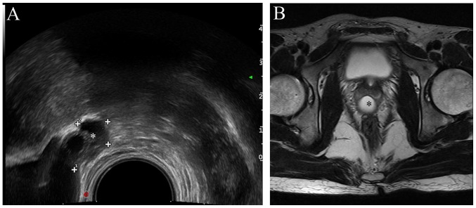
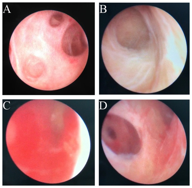
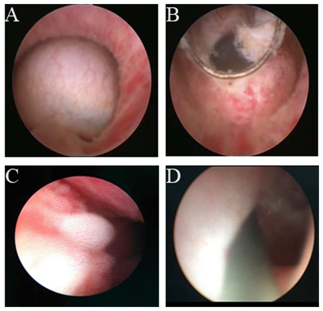
The major purpose of the present study was to investigate the efficacy and feasibility of the Mullerian duct cyst treatment by transurethral electrotomy combined with seminal vesiculoscopy. The clinical data of 20 aspermia patients who presented with Mullerian Cyst between March 2009 and March 2016 were retrospectively analyzed in the present study. Semen specimens of all patients were obtained by masturbation or sperm collector and diagnosed as aspermia by semen analysis (including sperm count, semen volume, sperm density, pH and fructose level). By transrectal ultrasonography, magnetic resonance imaging and testicular biopsy, the diagnosis of Mullerian cyst inducing obstruction aspermia was correctly identified. All patients were treated with the combination of transurethral resection and seminal vesiculoscopy. The operation time was 30-50 min. The follow-up duration after the operation was 12 months. All subjects included in the present study successfully underwent the operation. The semen quality of all patients was greatly improved and sperms were detected in semen specimens. The semen routine examination results of 3 consecutive follow-up exams within 12 months were within the normal range. The ejaculate volume and semen fructose levels were significantly higher than those prior to surgery (P<0.05). Furthermore, at 12 months post-operatively, the seminal vesicles of 6 patients were smaller than at the pre-operative stage. In conclusion, transurethral resection combined with seminal vesiculoscopy may be an effective and feasible option for the treatment of patients with Mullerian duct cyst.
本研究的主要目的是探讨经尿道电切联合精囊镜治疗苗勒管囊肿的疗效和可行性。本研究回顾性分析了2009年3月至2016年3月间20例因苗勒管囊肿导致无精子症患者的临床资料。所有患者的精液标本均通过手淫或取精器获取,并经精液分析(包括精子计数、精液量、精子密度、pH值和果糖水平)诊断为无精子症。通过经直肠超声、磁共振成像和睾丸活检,正确诊断为苗勒管囊肿导致梗阻性无精子症。所有患者均接受经尿道切除术联合精囊镜治疗。手术时间为30 - 50分钟。术后随访时间为12个月。本研究纳入的所有受试者均成功接受了手术。所有患者的精液质量均有显著改善,精液标本中检测到精子。12个月内连续3次随访的精液常规检查结果均在正常范围内。射精量和精液果糖水平显著高于术前(P<0.05)。此外,术后12个月时,6例患者的精囊较术前变小。总之,经尿道切除术联合精囊镜可能是治疗苗勒管囊肿患者的一种有效且可行的选择。