Li Jinghui, Zhang Fangming, Zhang Ning, Geng Xuefei, Meng Cen, Wang Xiaoying, Yang Ying
Department of stomatology, Beijing Friendship Hospital, Capital Medical University, Beijing, China.
PeerJ. 2019 Mar 8;7:e6589. doi: 10.7717/peerj.6589. eCollection 2019.
The periodontal ligament cells (PDLCs) contain heterogeneous cell populations and possess stem-cell-like properties. PDLCs have attracted considerable attention as an option for periodontal regeneration. However, the osteogenic differentiation of PDLCs remains obscure owing to variable osteo-inductive methods and whether PDLCs could be directly used for periodontal regeneration without stem cell enrichment is uncertain. The aim of the present study was to clarify the osteogenic differentiation capacity of PDLCs and test PDLCs as an alternative to stem cells for periodontal regeneration.
We tested the performance of human PDLCs in osteo-inductive culture and transplantation in vivo while taking human bone marrow derived mesenchymal stem cells (hMSCs) as positive control. Proliferation of PDLCs and hMSCs in osteo-inductive condition were examined by MTT assay and colony formation assay. The osteogenic differentiations of PDLCs and hMSCs were assessed by Alkaline phosphatase (ALP) activity measurement, von Kossa staining, Alizarin red S staining and quantitative RT-PCR of osteogenic marker gene including RUNX2, ALP, OCN, Col I, BSP, OPN. We transplanted osteo-inductive PDLCs and hMSCs with hydroxyapatite/tricalcium phosphate (HA/TCP) scaffolds to immunodeficient mice to explore their biological behaviors in vivo by histological staining and immunohistochemical evaluation.
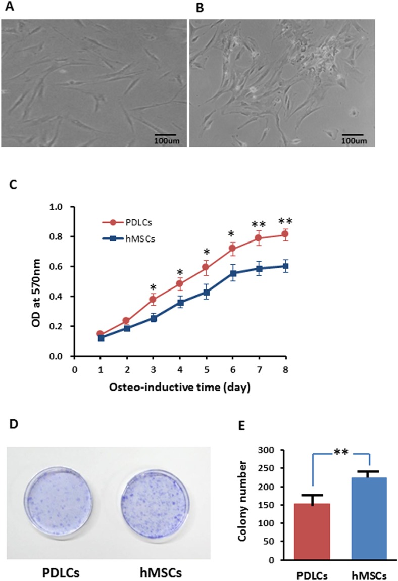
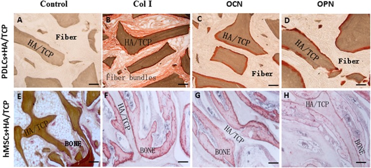
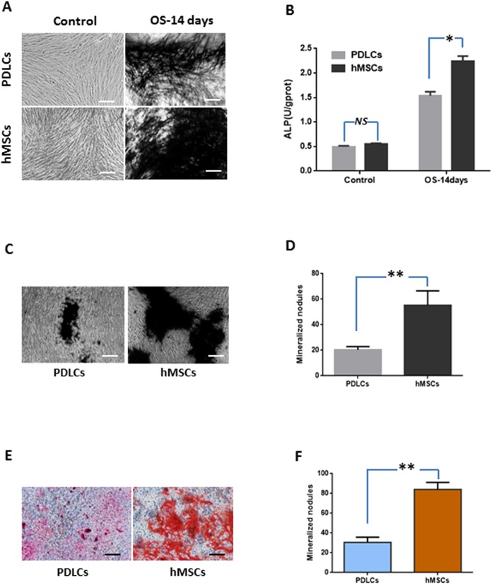
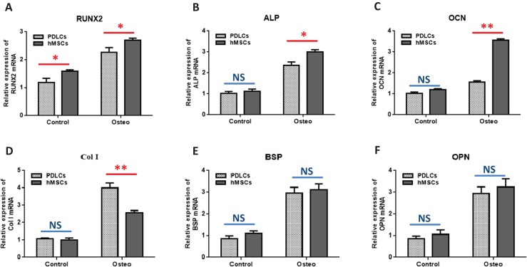
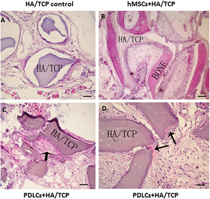
After 14 days of osteo-induction, PDLCs exhibited significantly higher proliferation rate but lower colony-forming ability comparing with hMSCs. PDLCs demonstrated lower ALP activity and generated fewer mineralized nodules than hMSCs. PDLCs showed overall up-regulated expression of RUNX2, ALP, OCN, Col I, BSP, OPN after osteo-induction. Col I level of PDLCs in osteo-inductive group was significantly higher while RUNX2, ALP, OCN were lower than that of hMSCs. Massive fiber bundles were produced linking or circling the scaffold while the bone-like structures were limited in the PDLCs-loaded HA/TCP samples. The fiber bundles displayed strong positive Col I, but weak OCN and OPN staining. The in vivo results were consistent with the in vitro data, which confirmed strong collagen forming ability and considerable osteogenic potential of PDLCs.
It is encouraging to find that PDLCs exhibit higher proliferation, stronger collagen fiber formation capacity, but lower osteogenic differentiation ability in comparison with hMSCs. This characteristic is essential for the successful periodontal reconstruction which is based on the synchronization of fiber formation and bone deposition. Moreover, PDLCs have advantages such as good accessibility, abundant source, vigorous proliferation and evident osteogenic differentiation capacity when triggered properly. They can independently form PDL-like structure in vivo without specific stem cell enrichment procedure. The application of PDLCs may offer a novel cytotherapeutic option for future clinical periodontal reconstruction.
牙周膜细胞(PDLCs)包含异质性细胞群体,并具有干细胞样特性。作为牙周再生的一种选择,PDLCs已引起了相当大的关注。然而,由于骨诱导方法的差异以及PDLCs能否不经干细胞富集而直接用于牙周再生尚不确定,PDLCs的成骨分化仍不清楚。本研究的目的是阐明PDLCs的成骨分化能力,并测试PDLCs作为干细胞替代物用于牙周再生的效果。
我们以人骨髓间充质干细胞(hMSCs)作为阳性对照,测试了人PDLCs在骨诱导培养和体内移植中的性能。通过MTT法和集落形成试验检测PDLCs和hMSCs在骨诱导条件下的增殖情况。通过碱性磷酸酶(ALP)活性测定、冯库萨染色、茜素红S染色以及对包括RUNX2、ALP、OCN、Col I、BSP、OPN在内的成骨标记基因进行定量RT-PCR,评估PDLCs和hMSCs的成骨分化情况。我们将经骨诱导的PDLCs和hMSCs与羟基磷灰石/磷酸三钙(HA/TCP)支架一起移植到免疫缺陷小鼠体内,通过组织学染色和免疫组化评估来探索它们在体内的生物学行为。
骨诱导14天后,与hMSCs相比,PDLCs表现出显著更高的增殖率,但集落形成能力较低。PDLCs的ALP活性较低,形成的矿化结节比hMSCs少。骨诱导后,PDLCs中RUNX2、ALP、OCN、Col I、BSP、OPN的表达总体上调。骨诱导组中PDLCs的Col I水平显著高于hMSCs,而RUNX2、ALP、OCN则低于hMSCs。在负载PDLCs的HA/TCP样本中,产生了大量连接或环绕支架的纤维束,而类骨结构有限。纤维束显示出强烈的Col I阳性,但OCN和OPN染色较弱。体内结果与体外数据一致,证实了PDLCs具有较强的胶原形成能力和相当的成骨潜力。
令人鼓舞的是,与hMSCs相比,PDLCs表现出更高的增殖能力、更强的胶原纤维形成能力,但成骨分化能力较低。这一特性对于基于纤维形成和骨沉积同步的成功牙周重建至关重要。此外,PDLCs具有易于获取、来源丰富、增殖旺盛以及在适当触发时具有明显成骨分化能力等优点。它们无需特定的干细胞富集程序就能在体内独立形成PDL样结构。PDLCs的应用可能为未来临床牙周重建提供一种新的细胞治疗选择。