• 文献检索
  • 文档翻译
  • 深度研究
  • 学术资讯
  • Suppr Zotero 插件Zotero 插件
  • 邀请有礼
  • 套餐&价格
  • 历史记录
应用&插件
Suppr Zotero 插件Zotero 插件浏览器插件Mac 客户端Windows 客户端微信小程序
定价
高级版会员购买积分包购买API积分包
服务
文献检索文档翻译深度研究API 文档MCP 服务
关于我们
关于 Suppr公司介绍联系我们用户协议隐私条款
关注我们

Suppr 超能文献

核心技术专利:CN118964589B侵权必究
粤ICP备2023148730 号-1Suppr @ 2026

文献检索

告别复杂PubMed语法,用中文像聊天一样搜索,搜遍4000万医学文献。AI智能推荐,让科研检索更轻松。

立即免费搜索

文件翻译

保留排版,准确专业,支持PDF/Word/PPT等文件格式,支持 12+语言互译。

免费翻译文档

深度研究

AI帮你快速写综述,25分钟生成高质量综述,智能提取关键信息,辅助科研写作。

立即免费体验

颅神经成像:图文概述。

Imaging of cranial nerves: a pictorial overview.

作者信息

Romano Nicola, Federici Margherita, Castaldi Antonio

机构信息

Department of Health Sciences (DISSAL) - Radiology Section, University of Genoa, Genoa, Italy.

Department of Diagnostic and Interventional Neuroradiology, E.O. Ospedali Galliera, Genoa, Italy.

出版信息

Insights Imaging. 2019 Mar 15;10(1):33. doi: 10.1186/s13244-019-0719-5.

DOI:10.1186/s13244-019-0719-5
PMID:30877408
原文链接:https://pmc.ncbi.nlm.nih.gov/articles/PMC6420596/
Abstract

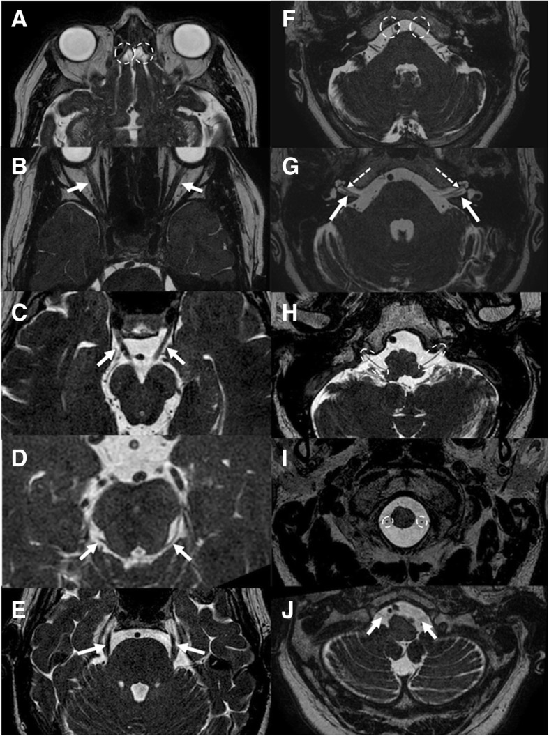
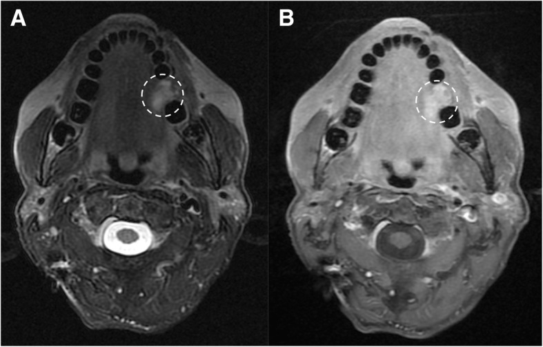
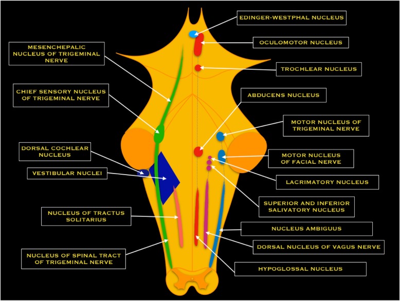
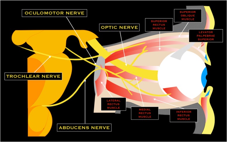
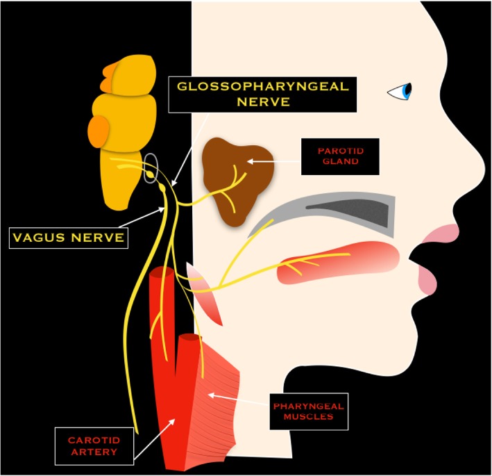
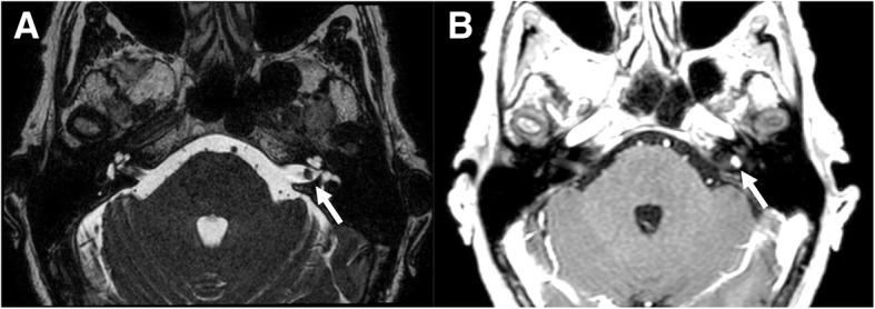
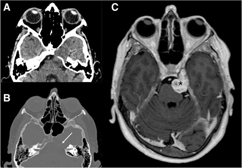
The human body has 12 pairs of cranial nerves that control motor and sensory functions of the head and neck. The anatomy of cranial nerves is complex and its knowledge is crucial to detect pathological alterations in case of nervous disorders. Therefore, it is necessary to know the most frequent pathologies that may involve cranial nerves and recognize their typical characteristics of imaging. Cranial nerve dysfunctions may be the result of pathological processes of the cranial nerve itself or be related to tumors, inflammation, infectious processes, or traumatic injuries of adjacent structures. Magnetic resonance imaging (MRI) is considered the gold standard in the study of the cranial nerves. Computed tomography (CT) allows, usually, an indirect view of the nerve and is useful to demonstrate the intraosseous segments of cranial nerves, the foramina through which they exit skull base and their pathologic changes. The article is a complete pictorial overview of the imaging of cranial nerves, with anatomic and pathologic descriptions and great attention to illustrative depiction. We believe that it could be a useful guide for radiologists and neuroradiologists to review the anatomy and the most important pathologies that involve cranial nerves and their differential diagnosis.

摘要

人体有12对颅神经,控制头部和颈部的运动及感觉功能。颅神经的解剖结构复杂,了解其知识对于在神经疾病情况下检测病理改变至关重要。因此,有必要了解可能累及颅神经的最常见病理情况,并认识其典型的影像学特征。颅神经功能障碍可能是颅神经本身病理过程的结果,也可能与肿瘤、炎症、感染性疾病或相邻结构的创伤性损伤有关。磁共振成像(MRI)被认为是研究颅神经的金标准。计算机断层扫描(CT)通常能间接观察神经,有助于显示颅神经的骨内段、它们穿出颅底的孔道及其病理变化。本文是颅神经影像学的完整图像综述,包括解剖和病理描述,并特别注重图示说明。我们认为,它可能是放射科医生和神经放射科医生回顾涉及颅神经的解剖结构、最重要的病理情况及其鉴别诊断的有用指南。

https://cdn.ncbi.nlm.nih.gov/pmc/blobs/a56f/6420596/0d4a856fa2a4/13244_2019_719_Fig15_HTML.jpg
https://cdn.ncbi.nlm.nih.gov/pmc/blobs/a56f/6420596/b5cbbd8dd3a7/13244_2019_719_Fig1_HTML.jpg
https://cdn.ncbi.nlm.nih.gov/pmc/blobs/a56f/6420596/2e9d6c76e7ce/13244_2019_719_Fig3_HTML.jpg
https://cdn.ncbi.nlm.nih.gov/pmc/blobs/a56f/6420596/b6da766035e7/13244_2019_719_Fig5_HTML.jpg
https://cdn.ncbi.nlm.nih.gov/pmc/blobs/a56f/6420596/67372d5e5a59/13244_2019_719_Fig7_HTML.jpg
https://cdn.ncbi.nlm.nih.gov/pmc/blobs/a56f/6420596/88ae55b76b3d/13244_2019_719_Fig9_HTML.jpg
https://cdn.ncbi.nlm.nih.gov/pmc/blobs/a56f/6420596/c6d537f92fe0/13244_2019_719_Fig11_HTML.jpg
https://cdn.ncbi.nlm.nih.gov/pmc/blobs/a56f/6420596/f4399f8b8a25/13244_2019_719_Fig13_HTML.jpg
https://cdn.ncbi.nlm.nih.gov/pmc/blobs/a56f/6420596/0d4a856fa2a4/13244_2019_719_Fig15_HTML.jpg

相似文献

1
Imaging of cranial nerves: a pictorial overview.颅神经成像:图文概述。
Insights Imaging. 2019 Mar 15;10(1):33. doi: 10.1186/s13244-019-0719-5.
2
ANATOMICAL STUDY OF CRANIAL NERVE EMERGENCE AND SKULL FORAMINA IN THE HORSE USING MAGNETIC RESONANCE IMAGING AND COMPUTED TOMOGRAPHY.利用磁共振成像和计算机断层扫描对马的颅神经出颅及颅骨孔道进行解剖学研究。
Vet Radiol Ultrasound. 2015 Jul-Aug;56(4):391-7. doi: 10.1111/vru.12256. Epub 2015 Apr 2.
3
3T MRI and 128-slice dual-source CT cisternography images of the cranial nerves a brief pictorial review for clinicians.3T磁共振成像和128层双源CT脑池造影术对颅神经成像:给临床医生的简要图像综述
Clin Anat. 2014 Jan;27(1):31-45. doi: 10.1002/ca.22311. Epub 2013 Dec 2.
4
Magnetic resonance imaging of the cranial nerves in infectious, neoplastic, and demyelinating diseases, as well as other inflammatory diseases: a pictorial essay.感染性、肿瘤性、脱髓鞘性疾病以及其他炎症性疾病中颅神经的磁共振成像:图文综述
Radiol Bras. 2022 Jan-Feb;55(1):38-46. doi: 10.1590/0100-3984.2021.0042.
5
[Imaging anatomy of the cranial nerves using 3.0 Tesla MRI: a practical review for clinicians].[使用3.0特斯拉磁共振成像对颅神经进行成像解剖:临床医生实用综述]
Gac Med Mex. 2011 Nov-Dec;147(6):526-37.
6
Magnetic resonance imaging of cranial nerves IX, X, XI, and XII.第九、十、十一和十二对颅神经的磁共振成像。
Top Magn Reson Imaging. 1996 Jun;8(3):180-6.
7
Cranial Nerve Disorders in Children: MR Imaging Findings.儿童颅神经疾病:磁共振成像表现
Radiographics. 2016 Jul-Aug;36(4):1178-94. doi: 10.1148/rg.2016150163.
8
Imaging the ocular motor nerves.眼运动神经影像学。
Eur J Radiol. 2010 May;74(2):314-22. doi: 10.1016/j.ejrad.2009.05.066. Epub 2010 Mar 11.
9
[Anatomy of the skull base and the cranial nerves in slice imaging].[切片成像中颅底及脑神经的解剖学]
Radiologe. 2009 Jul;49(7):584-97. doi: 10.1007/s00117-008-1800-0.
10
Magnetic resonance imaging of the cranial nerves in congenital, traumatic, and vascular diseases: a pictorial essay.先天性、创伤性和血管性疾病中颅神经的磁共振成像:图文综述
Radiol Bras. 2021 May-Jun;54(3):185-192. doi: 10.1590/0100-3984.2020.0039.

引用本文的文献

1
Imaging Findings in Neurogenic Ptosis.神经源性上睑下垂的影像学表现
J Korean Soc Radiol. 2025 Jul;86(4):483-500. doi: 10.3348/jksr.2024.0107. Epub 2025 Jul 25.
2
Challenges in Diagnosing the Course of the Lingual Nerve for Clinical Practice and Research.临床实践与研究中舌神经走行诊断的挑战
Diagnostics (Basel). 2025 Jun 25;15(13):1609. doi: 10.3390/diagnostics15131609.
3
Isolated abducens nerve involvement as a revealing sign of temporal bone fracture: A case report.孤立性展神经受累作为颞骨骨折的一个揭示性征象:一例报告。

本文引用的文献

1
MR Neurographic Evaluation of Facial and Neck Pain: Normal and Abnormal Craniospinal Nerves below the Skull Base.颅神经磁共振神经影像学评估:颅底以下头面部和颈部疼痛的正常和异常颅神经。
Radiographics. 2018 Sep-Oct;38(5):1498-1513. doi: 10.1148/rg.2018170194.
2
Most common sites on MRI of intracranial neoplastic leptomeningeal disease.颅内肿瘤性软脑膜疾病 MRI 最常见的部位。
J Clin Neurosci. 2017 Nov;45:252-256. doi: 10.1016/j.jocn.2017.07.020. Epub 2017 Aug 9.
3
A Case of Acute Vestibular Neuritis Visualized by Three-Dimensional FLAIR-VISTA Magnetic Resonance Imaging.
Radiol Case Rep. 2025 Jun 21;20(9):4483-4487. doi: 10.1016/j.radcr.2025.05.069. eCollection 2025 Sep.
4
Generation and characterization of BAC transgenic mouse lines expressing a fluorescent protein in trigeminal and dorsal root ganglion neurons.在三叉神经节和背根神经节神经元中表达荧光蛋白的BAC转基因小鼠品系的构建与鉴定
PLoS One. 2025 Jun 6;20(6):e0321014. doi: 10.1371/journal.pone.0321014. eCollection 2025.
5
Automatic segmentation and volume measurement of anterior visual pathway in brain 3D-T1WI using deep learning.基于深度学习的脑部3D-T1WI图像中视觉前通路的自动分割与体积测量
Front Med (Lausanne). 2025 Apr 28;12:1530361. doi: 10.3389/fmed.2025.1530361. eCollection 2025.
6
Blind spots in brain imaging: a pictorial essay.脑成像中的盲点:一篇图文并茂的文章。
Quant Imaging Med Surg. 2025 Jan 2;15(1):1023-1039. doi: 10.21037/qims-24-1270. Epub 2024 Dec 30.
7
Leontiasis ossea in the setting of chronic kidney disease and limited dialysis access: A case report with discussion of therapeutic implications.慢性肾脏病合并有限透析通路情况下的骨肥大症:一例报告并讨论治疗意义
Radiol Case Rep. 2024 Dec 12;20(3):1333-1337. doi: 10.1016/j.radcr.2024.11.049. eCollection 2025 Mar.
8
Health-Related Quality of Life with Iatrogenic Inferior Alveolar Nerve Injuries Treated with Photobiomodulation: A Comparative Study.光生物调节治疗医源性下牙槽神经损伤的健康相关生活质量:一项比较研究。
J Clin Med. 2024 Nov 28;13(23):7237. doi: 10.3390/jcm13237237.
9
On the Cranial Nerves.论颅神经。
NeuroSci. 2023 Dec 28;5(1):8-38. doi: 10.3390/neurosci5010002. eCollection 2024 Mar.
10
Characterizing neuroinvasion and neuropathology of SARS-CoV-2 by using AC70 human ACE2 transgenic mice.通过使用AC70人血管紧张素转换酶2转基因小鼠来表征新型冠状病毒的神经侵袭和神经病理学。
Front Microbiol. 2024 Sep 24;15:1455462. doi: 10.3389/fmicb.2024.1455462. eCollection 2024.
一例通过三维液体衰减反转恢复快速成像稳态进动(FLAIR-VISTA)磁共振成像可视化的急性前庭神经炎病例
Neuroophthalmology. 2014 Feb 25;38(2):60-61. doi: 10.3109/01658107.2013.874454. eCollection 2014.
4
Clinical-Radiologic Correlation of Extraocular Eye Movement Disorders: Seeing beneath the Surface.眼球运动障碍的临床与放射学关联:洞察表象之下
Radiographics. 2016 Nov-Dec;36(7):2123-2139. doi: 10.1148/rg.2016150227.
5
Cranial Nerve Schwannomas: Diagnostic Imaging Approach.颅神经鞘瘤:诊断成像方法。
Radiographics. 2016 Sep-Oct;36(5):1463-77. doi: 10.1148/rg.2016150199. Epub 2016 Aug 19.
6
Visualization of the olfactory nerve using constructive interference in steady state magnetic resonance imaging.利用稳态磁共振成像中的相长干涉对嗅神经进行可视化。
Surg Radiol Anat. 2017 Mar;39(3):315-321. doi: 10.1007/s00276-016-1731-9. Epub 2016 Aug 9.
7
Pathology of Perineural Spread.神经周围扩散的病理学
J Neurol Surg B Skull Base. 2016 Apr;77(2):124-30. doi: 10.1055/s-0036-1571837. Epub 2016 Feb 26.
8
Imaging Characteristics of Olfactory Neuroblastoma (Esthesioneuroblastoma).嗅神经母细胞瘤(嗅神经上皮瘤)的影像学特征
J Neurol Surg B Skull Base. 2016 Feb;77(1):1-5. doi: 10.1055/s-0035-1564053. Epub 2015 Sep 14.
9
Imaging of Neurovascular Compression Syndromes: Trigeminal Neuralgia, Hemifacial Spasm, Vestibular Paroxysmia, and Glossopharyngeal Neuralgia.神经血管压迫综合征的影像学表现:三叉神经痛、面肌痉挛、前庭阵发性眩晕和舌咽神经痛。
AJNR Am J Neuroradiol. 2016 Aug;37(8):1384-92. doi: 10.3174/ajnr.A4683. Epub 2016 Feb 18.
10
Magnetic resonance imaging of optic nerve.视神经的磁共振成像
Indian J Radiol Imaging. 2015 Oct-Dec;25(4):421-38. doi: 10.4103/0971-3026.169462.