• 文献检索
  • 文档翻译
  • 深度研究
  • 学术资讯
  • Suppr Zotero 插件Zotero 插件
  • 邀请有礼
  • 套餐&价格
  • 历史记录
应用&插件
Suppr Zotero 插件Zotero 插件浏览器插件Mac 客户端Windows 客户端微信小程序
定价
高级版会员购买积分包购买API积分包
服务
文献检索文档翻译深度研究API 文档MCP 服务
关于我们
关于 Suppr公司介绍联系我们用户协议隐私条款
关注我们

Suppr 超能文献

核心技术专利:CN118964589B侵权必究
粤ICP备2023148730 号-1Suppr @ 2026

文献检索

告别复杂PubMed语法,用中文像聊天一样搜索,搜遍4000万医学文献。AI智能推荐,让科研检索更轻松。

立即免费搜索

文件翻译

保留排版,准确专业,支持PDF/Word/PPT等文件格式,支持 12+语言互译。

免费翻译文档

深度研究

AI帮你快速写综述,25分钟生成高质量综述,智能提取关键信息,辅助科研写作。

立即免费体验

颈椎计算机断层血管摄影术上的点状钙与缺血性中风风险增加相关。

Spotty Calcium on Cervicocerebral Computed Tomography Angiography Associates With Increased Risk of Ischemic Stroke.

机构信息

From the Department of Radiology, Hainan Branch of Chinese People's Liberation Army General Hospital, Sanya, Hainan, China (F.Z.).

Biomedical Imaging Research Institute, Cedars-Sinai Medical Center, Los Angeles, CA (F.Z., Z.F., B.Z., Z.D., D.D., D.S.B., D.L., Y.X.).

出版信息

Stroke. 2019 Apr;50(4):859-866. doi: 10.1161/STROKEAHA.118.023273.

DOI:10.1161/STROKEAHA.118.023273
PMID:30879439
原文链接:https://pmc.ncbi.nlm.nih.gov/articles/PMC6433492/
Abstract

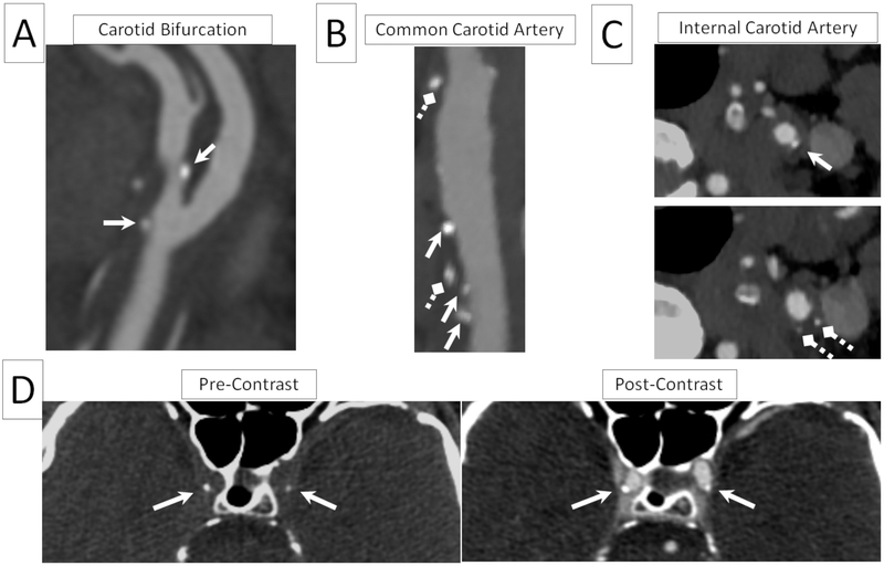
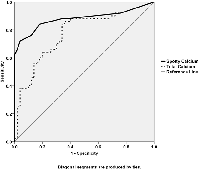
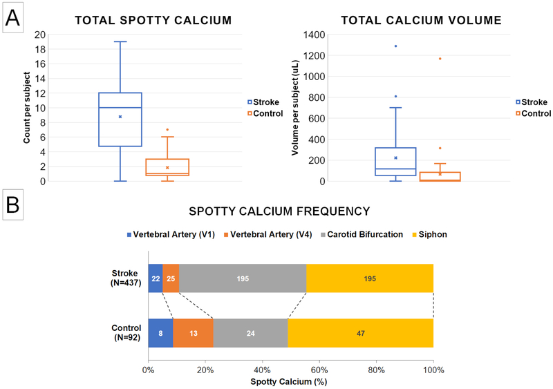
Background and Purpose- Cervicocerebral vascular calcification on computed tomography angiography is a known sign of advanced atherosclerosis. However, the clinical significance of calcification pattern remains unclear. In this study, we aimed to investigate the potential association between spotty calcium and acute ischemic stroke. Methods- This study included patients with first-time nonlacunar ischemic stroke (N=50) confirmed by brain magnetic resonance imaging or nonenhanced head computed tomography, as well as control subjects with asymptomatic carotid atherosclerosis (N=50) confirmed by carotid ultrasonography. Subjects in both groups underwent contrast-enhanced cervicocerebral computed tomography angiography within a week after the initial imaging examination. Spotty calcification was evaluated at 11 arterial segments commonly affected by atherosclerosis along the carotid and vertebrobasilar circulation. Statistical analysis was performed comparing the frequency and spatial pattern of spotty calcification between the 2 groups. Results- Spotty calcification in the Stroke group was markedly more prevalent than that in the Control group (total SC count: 8.74±4.96 versus 1.84±1.82, P<0.001). The odds ratio (95% CI) for stroke was 2.49 (1.55-4.00) for spotty calcification at bilateral carotid bifurcation, 1.52 (1.13-2.04) at carotid siphon, and 1.98 (1.45-2.69) at all evaluated locations. A total number of 3 spotty calcifications were determined as the optimal cutoff threshold for increased risk of stroke. Spotty calcium showed significantly greater area under the receiver operating characteristics curve than total calcium volume irrespective of size (0.88 versus 0.77). Within the Stroke group, ipsilateral lateral side showed significantly more spotty calcium than the contralateral side (5.18±3.05 versus 3.56±2.67, P<0.001). Conclusions- Nonlacunar ischemia stroke was associated with markedly increased incidence of spotty calcification with a distinct spatial pattern on cervicocerebral computed tomography compared with subclinical atherosclerosis, suggesting the potential role of spotty calcification for improving the risk stratification for ischemic stroke.

摘要

背景与目的- 计算机断层血管造影(CTA)显示的颈脑血管钙化是动脉粥样硬化进展的已知标志。然而,钙化模式的临床意义尚不清楚。本研究旨在探讨点状钙与急性缺血性卒中之间的潜在关联。方法- 本研究纳入了经脑磁共振成像或非增强头部 CT 证实的首次非腔隙性缺血性卒中(N=50)患者,以及经颈动脉超声证实的无症状颈动脉粥样硬化(N=50)的对照组患者。两组患者均在首次影像学检查后一周内行颈脑血管 CTA 增强检查。在颈动脉和椎基底动脉循环的 11 个常受动脉粥样硬化影响的动脉段评估点状钙化。对两组间点状钙化的频率和空间分布进行统计分析。结果- 卒中组的点状钙化明显比对照组更常见(总 SC 计数:8.74±4.96 对 1.84±1.82,P<0.001)。点状钙化双侧颈动脉分叉处、颈动脉虹吸处和所有评估部位的卒中比值比(95%可信区间)分别为 2.49(1.55-4.00)、1.52(1.13-2.04)和 1.98(1.45-2.69)。点状钙化 3 个被确定为卒中风险增加的最佳截断阈值。无论大小,点状钙的曲线下面积均明显大于总钙体积(0.88 对 0.77)。在卒中组中,同侧外侧比对侧侧显示出明显更多的点状钙化(5.18±3.05 对 3.56±2.67,P<0.001)。结论- 与亚临床动脉粥样硬化相比,非腔隙性缺血性卒中与颈脑血管 CTA 上点状钙化明显增加且具有明显的空间模式相关,表明点状钙化可能有助于改善缺血性卒中的风险分层。

https://cdn.ncbi.nlm.nih.gov/pmc/blobs/dc62/6433492/a98293cce07e/nihms-1521768-f0003.jpg
https://cdn.ncbi.nlm.nih.gov/pmc/blobs/dc62/6433492/1095111e1045/nihms-1521768-f0001.jpg
https://cdn.ncbi.nlm.nih.gov/pmc/blobs/dc62/6433492/646c50240cad/nihms-1521768-f0002.jpg
https://cdn.ncbi.nlm.nih.gov/pmc/blobs/dc62/6433492/a98293cce07e/nihms-1521768-f0003.jpg
https://cdn.ncbi.nlm.nih.gov/pmc/blobs/dc62/6433492/1095111e1045/nihms-1521768-f0001.jpg
https://cdn.ncbi.nlm.nih.gov/pmc/blobs/dc62/6433492/646c50240cad/nihms-1521768-f0002.jpg
https://cdn.ncbi.nlm.nih.gov/pmc/blobs/dc62/6433492/a98293cce07e/nihms-1521768-f0003.jpg

相似文献

1
Spotty Calcium on Cervicocerebral Computed Tomography Angiography Associates With Increased Risk of Ischemic Stroke.颈椎计算机断层血管摄影术上的点状钙与缺血性中风风险增加相关。
Stroke. 2019 Apr;50(4):859-866. doi: 10.1161/STROKEAHA.118.023273.
2
Prevalence of Ipsilateral Nonstenotic Carotid Plaques on Computed Tomography Angiography in Embolic Stroke of Undetermined Source.不明来源栓塞性卒中患者计算机断层血管造影术同侧非狭窄性颈动脉斑块的患病率。
Stroke. 2020 Jun;51(6):1743-1749. doi: 10.1161/STROKEAHA.120.029404. Epub 2020 May 7.
3
Increased incidence of napkin-ring sign plaques on cervicocerebral computed tomography angiography associated with the risk of acute ischemic stroke occurrence.颈脑计算机断层扫描血管造影上餐巾环征斑块的发生率增加与急性缺血性卒中发生风险相关。
Eur Radiol. 2024 Jul;34(7):4438-4447. doi: 10.1007/s00330-023-10404-w. Epub 2023 Nov 25.
4
Association Between Spotty Calcification in Nonstenosing Extracranial Carotid Artery Plaque and Ipsilateral Ischemic Stroke.非狭窄性颅外颈动脉斑块内斑点状钙化与同侧缺血性卒中的关系。
J Am Heart Assoc. 2023 May 16;12(10):e028525. doi: 10.1161/JAHA.122.028525. Epub 2023 May 15.
5
Extracranial Carotid Plaque Calcification and Cerebrovascular Ischemia: A Systematic Review and Meta-Analysis.颅外颈动脉斑块钙化与脑血管缺血:系统评价和荟萃分析。
Stroke. 2023 Oct;54(10):2621-2628. doi: 10.1161/STROKEAHA.123.042807. Epub 2023 Aug 28.
6
Serum Aldosterone Is Associated with Cerebral Artery Atherosclerosis and Calcification.血清醛固酮与脑动脉粥样硬化及钙化相关。
J Stroke Cerebrovasc Dis. 2019 Mar;28(3):523-530. doi: 10.1016/j.jstrokecerebrovasdis.2018.09.053. Epub 2018 Dec 8.
7
Letter by Ni et al Regarding Article "Spotty Calcium on Cervicocerebral Computed Tomography Angiography Associates with Increased Risk of Ischemic Stroke".倪等人就“颈脑计算机断层扫描血管造影上的斑点状钙化与缺血性中风风险增加相关”一文的信函
Stroke. 2019 Aug;50(8):e231. doi: 10.1161/STROKEAHA.119.025913. Epub 2019 Jun 27.
8
Response by Zhang and Xie to Letter Regarding Article, "Spotty Calcium on Cervicocerebral Computed Tomography Angiography Associates With Increased Risk of Ischemic Stroke".张和谢对关于文章《颈脑计算机断层扫描血管造影上的斑点状钙化与缺血性中风风险增加相关》的信件的回应。
Stroke. 2019 Aug;50(8):e232. doi: 10.1161/STROKEAHA.119.026032. Epub 2019 Jun 27.
9
Computed tomography evaluation of intracranial vascular calcification in major ischemic stroke patients (vascular territory)--its distribution and association with vascular risk factors: a retrospective trial.主要缺血性脑卒中患者颅内血管钙化的计算机断层扫描评估(血管区域)——其分布及与血管危险因素的关联:一项回顾性试验
J Med Assoc Thai. 2015 Apr;98(4):414-22.
10
Association between arterial calcifications and nonlacunar and lacunar ischemic strokes.动脉钙化与非腔隙性和腔隙性缺血性卒中的关系。
Stroke. 2014 Mar;45(3):728-33. doi: 10.1161/STROKEAHA.113.003197. Epub 2014 Jan 23.

引用本文的文献

1
Paradox of Exercise and Coronary Artery Calcification: Potential Underlying Mechanisms.运动与冠状动脉钙化的悖论:潜在的潜在机制。
Circ Res. 2025 Jul 7;137(2):335-349. doi: 10.1161/CIRCRESAHA.125.326011. Epub 2025 Jul 3.
2
Association between vertebrobasilar artery calcification and artery-to-artery embolism in the posterior circulation.椎基底动脉钙化与后循环动脉到动脉栓塞之间的关联。
Sci Rep. 2025 Jul 2;15(1):22689. doi: 10.1038/s41598-025-07229-6.
3
A Case of Asymptomatic Calcified Cerebral Emboli Harboring a Vulnerable Calcified Carotid Plaque: Diagnostic and Therapeutic Considerations.

本文引用的文献

1
Association between Intracranial Atherosclerotic Calcium Burden and Angiographic Luminal Stenosis Measurements.颅内动脉粥样硬化钙负荷与血管造影管腔狭窄测量的相关性。
AJNR Am J Neuroradiol. 2017 Sep;38(9):1723-1729. doi: 10.3174/ajnr.A5310. Epub 2017 Jul 20.
2
Cryptogenic Stroke and Nonstenosing Intracranial Calcified Atherosclerosis.隐源性卒中与非狭窄性颅内钙化性动脉粥样硬化
J Stroke Cerebrovasc Dis. 2017 Apr;26(4):863-870. doi: 10.1016/j.jstrokecerebrovasdis.2016.10.035. Epub 2016 Nov 23.
3
Intracranial Artery Calcification and Its Clinical Significance.
一例无症状性钙化脑栓塞合并易损钙化颈动脉斑块病例:诊断与治疗考量
Cureus. 2025 May 26;17(5):e84860. doi: 10.7759/cureus.84860. eCollection 2025 May.
4
In-Depth Carotid Calcification Morphometrics and Their Temporal Changes Are Associated with Cardiovascular Risk Factors in Patients with Recent Ischemic Event: The Plaque At Risk Study.深度颈动脉钙化形态计量学及其时间变化与近期缺血性事件患者的心血管危险因素相关:危险斑块研究
Cerebrovasc Dis. 2025 May 2:1-11. doi: 10.1159/000546164.
5
Extracranial carotid plaque calcification and its association with risk factors for cerebrovascular events: insights from the ANTIQUE study.颅外颈动脉斑块钙化及其与脑血管事件危险因素的关联:来自ANTIQUE研究的见解。
Front Neurol. 2025 Jan 29;16:1532883. doi: 10.3389/fneur.2025.1532883. eCollection 2025.
6
High-risk plaque characteristics associated with recurrent stroke in patients with intracranial stenosis: a systematic review and meta-analysis.颅内狭窄患者复发性卒中相关的高危斑块特征:一项系统评价和荟萃分析。
J Neurol. 2025 Feb 1;272(2):173. doi: 10.1007/s00415-025-12924-5.
7
Association between atherosclerosis and the development of multi-organ pathologies.动脉粥样硬化与多器官病变发展之间的关联。
SAGE Open Med. 2024 Dec 23;12:20503121241310013. doi: 10.1177/20503121241310013. eCollection 2024.
8
Calcified cerebral emboli associated with calcified carotid plaque: a case report and morphological consideration for plaque calcification.与颈动脉斑块钙化相关的钙化性脑栓塞:病例报告和斑块钙化的形态学考虑。
Acta Neurochir (Wien). 2024 Nov 4;166(1):439. doi: 10.1007/s00701-024-06339-w.
9
Prevalence and clinical implications of calcification in internal carotid artery stenosis: a retrospective study.颈内动脉狭窄中钙化的流行情况及其临床意义:一项回顾性研究。
BMC Neurol. 2024 Aug 10;24(1):279. doi: 10.1186/s12883-024-03788-9.
10
Precranial artery calcification burden: a potential indicator of the clinical outcome of reperfusion in patients with acute large artery occlusion.颅前动脉钙化负担:急性大动脉闭塞患者再灌注临床结局的潜在指标。
J Int Med Res. 2024 Jul;52(7):3000605241260364. doi: 10.1177/03000605241260364.
颅内动脉钙化及其临床意义
J Clin Neurol. 2016 Jul;12(3):253-61. doi: 10.3988/jcn.2016.12.3.253. Epub 2016 May 10.
4
Defining Core and Penumbra in Ischemic Stroke: A Voxel- and Volume-Based Analysis of Whole Brain CT Perfusion.缺血性卒中核心区和半暗带的定义:基于体素和体积的全脑CT灌注分析
Sci Rep. 2016 Feb 10;6:20932. doi: 10.1038/srep20932.
5
Intracranial Carotid Calcification on Cranial Computed Tomography: Visual Scoring Methods, Semiautomated Scores, and Volume Measurements in Patients With Stroke.头颅计算机断层扫描上的颅内颈动脉钙化:中风患者的视觉评分方法、半自动评分及体积测量
Stroke. 2015 Sep;46(9):2504-9. doi: 10.1161/STROKEAHA.115.009716. Epub 2015 Aug 6.
6
2015 American Heart Association/American Stroke Association Focused Update of the 2013 Guidelines for the Early Management of Patients With Acute Ischemic Stroke Regarding Endovascular Treatment: A Guideline for Healthcare Professionals From the American Heart Association/American Stroke Association.2015年美国心脏协会/美国卒中协会对2013年急性缺血性卒中患者早期管理指南中血管内治疗部分的重点更新:美国心脏协会/美国卒中协会给医疗专业人员的指南
Stroke. 2015 Oct;46(10):3020-35. doi: 10.1161/STR.0000000000000074. Epub 2015 Jun 29.
7
Impact of spotty calcification on long-term prediction of future revascularization: a prospective three-vessel intravascular ultrasound study.斑点状钙化对未来血运重建长期预测的影响:一项前瞻性三支血管血管内超声研究
Heart Vessels. 2016 Jun;31(6):881-9. doi: 10.1007/s00380-015-0687-8. Epub 2015 May 12.
8
Combining Coronary with Carotid and Cerebrovascular Angiography Using Prospective ECG Gating and Iterative Reconstruction with 256-slice CT.使用前瞻性心电图门控和256层CT迭代重建技术联合进行冠状动脉、颈动脉和脑血管造影
Echocardiography. 2015 Aug;32(8):1291-8. doi: 10.1111/echo.12830. Epub 2014 Nov 5.
9
Comparison of quantitative atherosclerotic plaque burden from coronary CT angiography in patients with first acute coronary syndrome and stable coronary artery disease.首次急性冠状动脉综合征患者与稳定型冠状动脉疾病患者冠状动脉CT血管造影定量动脉粥样硬化斑块负荷的比较。
J Cardiovasc Comput Tomogr. 2014 Sep-Oct;8(5):368-74. doi: 10.1016/j.jcct.2014.07.007. Epub 2014 Aug 23.
10
Interscan reproducibility of quantitative coronary plaque volume and composition from CT coronary angiography using an automated method.使用自动化方法对CT冠状动脉造影定量冠状动脉斑块体积和成分进行的两次扫描间的可重复性
Eur Radiol. 2014 Sep;24(9):2300-8. doi: 10.1007/s00330-014-3253-3. Epub 2014 Jun 25.