Neurologic Clinic and Policlinic (B.P., T.J.D., T.S.), Departments of Medicine, Clinical Research and Biomedical Engineering, University Hospital and University of Basel, Basel, Switzerland; NeuroCure Clinical Research Center (N.B., L.R., J.B.-S., F.P., T.S.), Charité-Universitätsmedizin Berlin, Corporate Member of Freie Universität Berlin, Humboldt-Universität zu Berlin, and Berlin Institute of Health; Department of Neurology (N.B., J.B.-S., K.R., F.P.), Charité-Universitätsmedizin Berlin, Corporate Member of Freie Universität Berlin, Humboldt-Universität zu Berlin, and Berlin Institute of Health; Experimental and Clinical Research Center (F.P.), Charité-Universitätsmedizin Berlin and Max Delbrück Center for Molecular Medicine; Clinical and Experimental Multiple Sclerosis Research Center (K.R., F.P.), Charité-Universitätsmedizin Berlin, Corporate Member of Freie Universität Berlin, Humboldt-Universität zu Berlin, and Berlin Institute of Health; Berlin Ultrahigh Field Facility (T.N.), Max Delbrück Center for Molecular Medicine in the Helmholtz Association, Germany; Medical Image Analysis Center AG (J.W., T.S.); and qbig (J.W.), Department of Biomedical Engineering, University of Basel, Basel, Switzerland.
Neurol Neuroimmunol Neuroinflamm. 2019 Mar 7;6(3):e541. doi: 10.1212/NXI.0000000000000541. eCollection 2019 May.
To investigate and compare occult damages in aquaporin-4 (AQP4)-rich periependymal regions in patients with neuromyelitis optica spectrum disorder (NMOSD) vs healthy controls (HCs) and patients with multiple sclerosis (MS) applying quantitative T1 mapping at 7 Tesla (T) in a cross-sectional study.
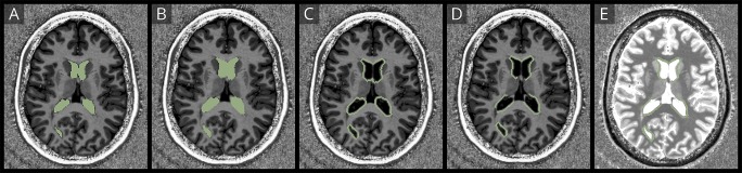
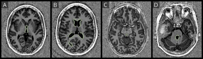
Eleven patients with NMOSD (median Expanded Disability Status Scale [EDSS] score 3.5, disease duration 9.3 years, age 43.7 years, and 11 female) seropositive for anti-AQP4 antibodies, 7 patients with MS (median EDSS score 1.5, disease duration 3.6, age 30.2 years, and 4 female), and 10 HCs underwent 7T MRI. The imaging protocol included T2*-weighted (w) imaging and an MP2RAGE sequence yielding 3D T1w images and quantitative T1 maps. We semiautomatically marked the lesion-free periependymal area around the cerebral aqueduct and the lateral, third, and fourth ventricles to finally measure and compare the T1 relaxation time within these areas.
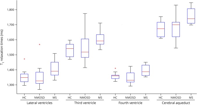
We did not observe any differences in the T1 relaxation time between patients with NMOSD and HCs (all > 0.05). Contrarily, the T1 relaxation time was longer in patients with MS vs patients with NMOSD (lateral ventricle = 0.056, third ventricle = 0.173, fourth ventricle = 0.016, and cerebral aqueduct = 0.048) and vs HCs (third ventricle = 0.027, fourth ventricle = 0.013, lateral ventricle = 0.043, and cerebral aqueduct = 0.005).
Unlike in MS, we did not observe subtle T1 changes in lesion-free periependymal regions in NMOSD, which supports the hypothesis of a rather focal than diffuse brain pathology in NMOSD.
在横断面研究中,通过 7 特斯拉(T)的定量 T1 映射,研究并比较视神经脊髓炎谱系障碍(NMOSD)患者与健康对照(HCs)和多发性硬化(MS)患者富含水通道蛋白 4(AQP4)的室管膜周围区域的隐匿性损伤。
11 例 NMOSD 患者(中位扩展残疾状况量表 [EDSS]评分 3.5,疾病病程 9.3 年,年龄 43.7 岁,女性 11 例)抗 AQP4 抗体阳性,7 例 MS 患者(中位 EDSS 评分 1.5,疾病病程 3.6 年,年龄 30.2 岁,女性 4 例)和 10 例 HCs 接受了 7T MRI 检查。成像方案包括 T2*-加权(w)成像和 MP2RAGE 序列,生成 3D T1w 图像和定量 T1 图谱。我们半自动化地标记了脑导水管周围和侧脑室、第三脑室和第四脑室周围无病变的室管膜区域,最终测量并比较了这些区域内的 T1 弛豫时间。
我们没有观察到 NMOSD 患者与 HCs 之间 T1 弛豫时间有任何差异(均>0.05)。相反,MS 患者的 T1 弛豫时间长于 NMOSD 患者(侧脑室=0.056,第三脑室=0.173,第四脑室=0.016,脑导水管=0.048)和 HCs(第三脑室=0.027,第四脑室=0.013,侧脑室=0.043,脑导水管=0.005)。
与 MS 不同,我们没有观察到 NMOSD 无病变室管膜周围区域的细微 T1 变化,这支持了 NMOSD 中脑病理学以局灶性而非弥漫性为主的假说。