Schmidt Felix A, Chien Claudia, Kuchling Joseph, Bellmann-Strobl Judith, Ruprecht Klemens, Siebert Nadja, Asseyer Susanna, Jarius Sven, Brandt Alexander U, Scheel Michael, Paul Friedemann
NeuroCure Clinical Research Center, Charité - Universitätsmedizin Berlin, Corporate Member of Freie Universität Berlin, Humboldt-Universität zu Berlin, and Berlin Institute of Health, Berlin, Germany.
Department of Neurology, Charité - Universitätsmedizin Berlin, Corporate Member of Freie Universität Berlin, Humboldt-Universität zu Berlin, and Berlin Institute of Health, Berlin, Germany.
Front Neurol. 2020 Sep 30;11:499910. doi: 10.3389/fneur.2020.499910. eCollection 2020.
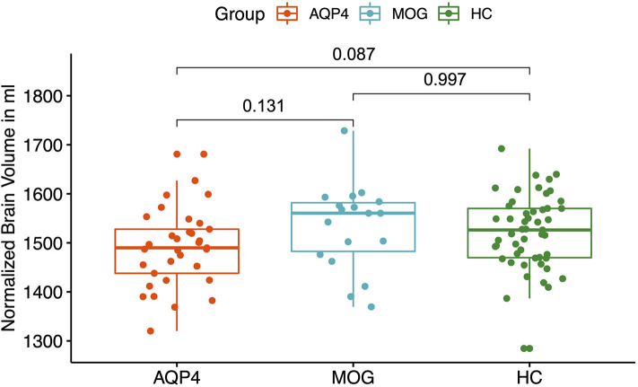
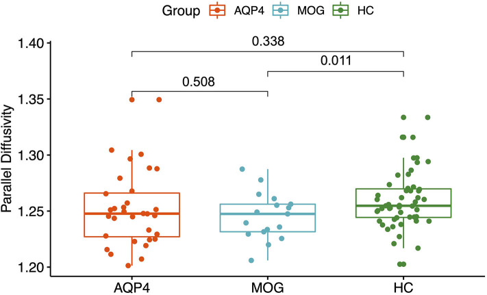
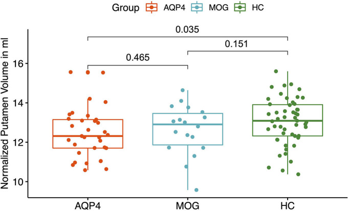
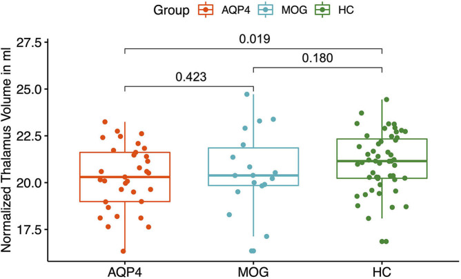
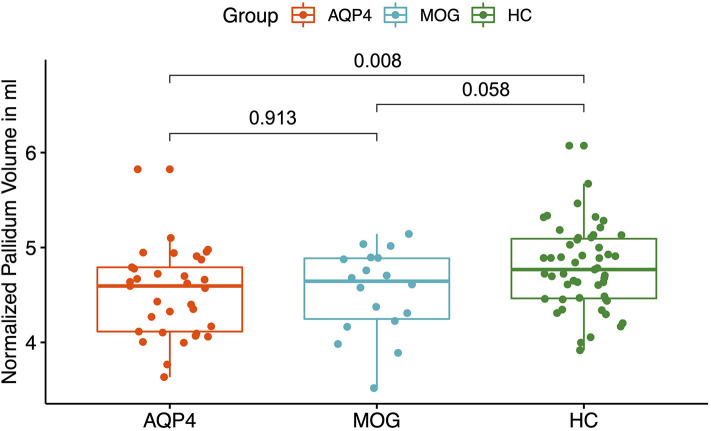
To explore differences in advanced brain magnetic resonance imaging (MRI) characteristics between myelin oligodendrocyte (MOG) immunoglobulin (IgG) and aquaporin-4 (AQP4) IgG seropositive (+) neuromyelitis optica spectrum disorders (NMOSD). 33 AQP4-IgG and 18 MOG-IgG seropositive NMOSD patients and 61 healthy control (HC) subjects were included. All 112 participants were scanned with the same standardized MRI-protocol on a 3-Tesla MRI-scanner. Brain volume and diffusion tensor imaging (DTI) parameters were assessed. MOG-IgG+ patients showed reduced parallel diffusivity within white matter tracts compared to HC whereas AQP4-IgG+ showed no significant brain parenchymal damage in DTI analysis. AQP4-IgG+ patients showed reduced whole brain volumes and reduced volumes of several deep gray matter structures compared to HC whereas MOG-IgG+ patients did not show reduced brain or deep gray matter volumes compared to HC. Microstructural brain parenchymal damage in MOG-IgG+ patients was more pronounced than in AQP4-IgG+ patients, compared with HC, whereas normalized brain volume reduction was more severe in AQP4-IgG+ patients. Longitudinal imaging studies are warranted to further investigate this trend in NMOSD. Our results suggest that MOG-IgG+ and AQP4-IgG+ NMOSD patients differ in cerebral MRI characteristics. Advanced MRI analysis did not help to differentiate between MOG-IgG+ and AQP4-IgG+ patients in our study.
为探究髓鞘少突胶质细胞(MOG)免疫球蛋白(IgG)和水通道蛋白4(AQP4)IgG血清阳性(+)的视神经脊髓炎谱系障碍(NMOSD)患者之间高级脑磁共振成像(MRI)特征的差异。纳入了33例AQP4-IgG血清阳性和18例MOG-IgG血清阳性的NMOSD患者以及61名健康对照(HC)受试者。所有112名参与者均在3特斯拉MRI扫描仪上采用相同的标准化MRI方案进行扫描。评估脑容量和扩散张量成像(DTI)参数。与HC相比,MOG-IgG+患者白质束内的平行扩散率降低,而在DTI分析中,AQP4-IgG+患者未显示明显的脑实质损伤。与HC相比,AQP4-IgG+患者全脑容量减小,几个深部灰质结构的体积也减小,而MOG-IgG+患者与HC相比未显示脑或深部灰质体积减小。与HC相比,MOG-IgG+患者的脑实质微观结构损伤比AQP4-IgG+患者更明显,而AQP4-IgG+患者的标准化脑容量减少更严重。有必要进行纵向成像研究以进一步探究NMOSD中的这一趋势。我们的结果表明,MOG-IgG+和AQP4-IgG+的NMOSD患者在脑MRI特征方面存在差异。在我们的研究中,高级MRI分析无助于区分MOG-IgG+和AQP4-IgG+患者。