Turku PET Centre, University of Turku, FI-20520 Turku, Finland.
Turku Center for Disease Modeling, University of Turku, FI-20520 Turku, Finland.
Molecules. 2019 Mar 19;24(6):1072. doi: 10.3390/molecules24061072.
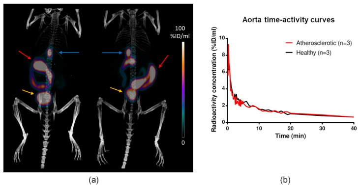
Atherosclerosis is characterized by the accumulation of oxidized lipids in the artery wall, which triggers an inflammatory response. Oxidized low-density lipoprotein (ox-LDL) presents amyloid-like structural properties, and different amyloid species have recently been recognized in atherosclerotic plaques. Therefore, we studied the uptake of the amyloid imaging agent [F]Flutemetamol in atherosclerotic plaques. The binding of [F]Flutemetamol to human carotid artery plaque was studied in vitro. In vivo uptake of the tracer was studied in hypercholesterolemic IGF-II/LDLRApoB mice and C57BL/6N controls. Tracer biodistribution was studied in vivo with PET/CT, and ex vivo by gamma counter and digital ex vivo autoradiography. The presence of amyloid, ox-LDL, and macrophages in the plaques was examined by immunohistochemistry. [F]Flutemetamol showed specific accumulation in human carotid plaque, especially in areas positive for amyloid beta. The aortas of IGF-II/LDLRApoB mice showed large thioflavin-S-positive atherosclerotic plaques containing ox-LDL and macrophages. Autoradiography revealed 1.7-fold higher uptake in the plaques than in a lesion-free vessel wall, but no difference in aortic tissue uptake between mouse strains were observed in the in vivo PET/CT. In conclusion, [F]Flutemetamol binds to amyloid-positive areas in human atherosclerotic plaques. Further studies are warranted to clarify the uptake mechanisms, and the potential of the tracer for in vivo imaging of atherosclerosis in patients.
动脉粥样硬化的特征是氧化脂质在动脉壁中的积累,这引发了炎症反应。氧化低密度脂蛋白(ox-LDL)呈现出类似淀粉样的结构特性,最近在动脉粥样硬化斑块中已经识别出不同的淀粉样物质。因此,我们研究了淀粉样成像剂[F]Flutemetamol在动脉粥样硬化斑块中的摄取。我们在体外研究了[F]Flutemetamol与人颈动脉斑块的结合。在高胆固醇血症 IGF-II/LDLRApoB 小鼠和 C57BL/6N 对照中研究了示踪剂的体内摄取。使用 PET/CT 研究了示踪剂的体内生物分布,并用伽马计数器和数字离体放射自显影术进行了离体研究。通过免疫组织化学检查斑块中淀粉样蛋白、ox-LDL 和巨噬细胞的存在。[F]Flutemetamol在人颈动脉斑块中表现出特异性积累,尤其是在淀粉样β阳性区域。IGF-II/LDLRApoB 小鼠的主动脉显示出大的硫黄素-S 阳性动脉粥样硬化斑块,含有 ox-LDL 和巨噬细胞。放射自显影显示斑块的摄取比无病变的血管壁高 1.7 倍,但在体内 PET/CT 中未观察到两种小鼠品系的主动脉组织摄取差异。总之,[F]Flutemetamol与人类动脉粥样硬化斑块中的淀粉样阳性区域结合。需要进一步的研究来阐明摄取机制,以及该示踪剂在患者体内动脉粥样硬化成像中的潜力。