• 文献检索
  • 文档翻译
  • 深度研究
  • 学术资讯
  • Suppr Zotero 插件Zotero 插件
  • 邀请有礼
  • 套餐&价格
  • 历史记录
应用&插件
Suppr Zotero 插件Zotero 插件浏览器插件Mac 客户端Windows 客户端微信小程序
定价
高级版会员购买积分包购买API积分包
服务
文献检索文档翻译深度研究API 文档MCP 服务
关于我们
关于 Suppr公司介绍联系我们用户协议隐私条款
关注我们

Suppr 超能文献

核心技术专利:CN118964589B侵权必究
粤ICP备2023148730 号-1Suppr @ 2026

文献检索

告别复杂PubMed语法,用中文像聊天一样搜索,搜遍4000万医学文献。AI智能推荐,让科研检索更轻松。

立即免费搜索

文件翻译

保留排版,准确专业,支持PDF/Word/PPT等文件格式,支持 12+语言互译。

免费翻译文档

深度研究

AI帮你快速写综述,25分钟生成高质量综述,智能提取关键信息,辅助科研写作。

立即免费体验

60例牙源性角化囊肿中的朗格汉斯细胞。

Langerhans cells in 60 odontogenic keratocysts.

作者信息

Chang Chun-Han, Wu Yang-Che, Wu Yu-Hsueh, Sun Andy, Chen Hsin-Ming, Lin Hung-Pin

机构信息

Graduate Institute of Oral Biology, School of Dentistry, National Taiwan University, Taipei, Taiwan.

Graduate Institute of Clinical Dentistry, School of Dentistry, National Taiwan University, Taipei, Taiwan.

出版信息

J Dent Sci. 2017 Sep;12(3):283-290. doi: 10.1016/j.jds.2017.04.001. Epub 2017 May 9.

DOI:10.1016/j.jds.2017.04.001
PMID:30895063
原文链接:https://pmc.ncbi.nlm.nih.gov/articles/PMC6399999/
Abstract

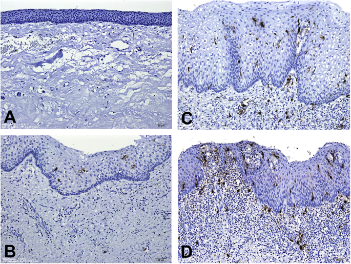
BACKGROUND/PURPOSE: Langerhans cells (LCs) are antigen-presenting cells. This study mainly evaluated the LC counts in 60 odontogenic keratocysts (OKCs).

MATERIALS AND METHODS

The CD1a-positive LC numbers in the lining epithelia and subepithelial connective tissues were counted at 60 OKC sites without inflammation, 39 OKC sites with mild/moderate inflammation, and 13 OKC sites with severe inflammation from 60 OKC specimens.

RESULTS

The mean CD1a-positive LC counts in the lining epithelia and subepithelial connective tissues increased significantly from no inflammation (0.5 ± 0.4 and 0.2 ± 0.3 cell/high-power field or HPF, respectively) through mild/moderate inflammation (5.3 ± 2.5 and 2.5 ± 2.7 cells/HPF, respectively) to severe inflammation OKC sites (12.7 ± 5.6 and 9.3 ± 7.2 cells/HPF, respectively; all P-values < 0.001). OKC sites with inflammation had thicker lining epithelia than those without inflammation. Moreover, the mean CD1a-positive LC counts in the lining epithelia and subepithelial connective tissues of OKCs were significantly higher in the thicker lining epithelium (>100 μm) group (6.8 ± 5.1 and 3.7 ± 4.9 cells/HPF, respectively) than in the thinner lining epithelium (≦100 μm) group (1.0 ± 1.7 and 0.8 ± 2.5 cell/HPF, respectively; both P-values < 0.001).

CONCLUSION

There is a significant association of inflammation grade with the number of LCs in OKCs. The scarce LCs in the lining epithelia of OKCs without inflammation suggests the loss of immunosurveillance ability against the OKC lining epithelial cells; this can explain why OKCs have aggressive clinical behavior, a great growth potential, and a high recurrence rate.

摘要

背景/目的:朗格汉斯细胞(LCs)是抗原呈递细胞。本研究主要评估60例牙源性角化囊肿(OKCs)中的LC计数。

材料与方法

从60例OKC标本中,对60个无炎症的OKC部位、39个有轻/中度炎症的OKC部位和13个有重度炎症的OKC部位的衬里上皮和上皮下结缔组织中的CD1a阳性LC数量进行计数。

结果

衬里上皮和上皮下结缔组织中CD1a阳性LC的平均计数从无炎症(分别为0.5±0.4和0.2±0.3个细胞/高倍视野或HPF)到轻/中度炎症(分别为5.3±2.5和2.5±2.7个细胞/HPF)再到重度炎症的OKC部位(分别为12.7±5.6和9.3±7.2个细胞/HPF)显著增加(所有P值<0.001)。有炎症的OKC部位的衬里上皮比无炎症的更厚。此外,OKC衬里上皮和上皮下结缔组织中CD1a阳性LC的平均计数在较厚衬里上皮(>100μm)组(分别为6.8±5.1和3.7±4.9个细胞/HPF)显著高于较薄衬里上皮(≤100μm)组(分别为1.0±1.7和0.8±2.5个细胞/HPF;两个P值<0.001)。

结论

炎症分级与OKC中LC的数量存在显著关联。无炎症的OKC衬里上皮中LC稀少表明对OKC衬里上皮细胞的免疫监视能力丧失;这可以解释为什么OKC具有侵袭性的临床行为、巨大的生长潜力和高复发率。

https://cdn.ncbi.nlm.nih.gov/pmc/blobs/7a17/6399999/3825b4835370/gr1.jpg

相似文献

1
Langerhans cells in 60 odontogenic keratocysts.60例牙源性角化囊肿中的朗格汉斯细胞。
J Dent Sci. 2017 Sep;12(3):283-290. doi: 10.1016/j.jds.2017.04.001. Epub 2017 May 9.
2
Significant association of inflammation grade with the number of Langerhans cells in odontogenic keratocysts.炎症程度与牙源性角化囊肿中朗格汉斯细胞数量存在显著相关性。
J Formos Med Assoc. 2017 Oct;116(10):798-805. doi: 10.1016/j.jfma.2017.04.002. Epub 2017 Jul 26.
3
Significant association of high-grade inflammation and thick lining epithelium with the increased number of Langerhans cells in dentigerous cysts.高等级炎症和厚衬里上皮与含牙囊肿中朗格汉斯细胞数量增加有显著关联。
J Formos Med Assoc. 2017 Nov;116(11):837-843. doi: 10.1016/j.jfma.2017.06.011. Epub 2017 Jul 24.
4
S100 protein-positive Langerhans cells in 80 dentigerous cysts.80例含牙囊肿中的S100蛋白阳性朗格汉斯细胞
J Dent Sci. 2017 Dec;12(4):405-412. doi: 10.1016/j.jds.2017.08.001. Epub 2017 Sep 25.
5
Sub-epithelial hyalinization, incomplete cystic lining, and corrugated surface could be a predictor of recurrence in Odontogenic Keratocysts.上皮下玻璃样变、囊壁内衬不全及表面呈波纹状可能是牙源性角化囊肿复发的一个预测指标。
J Oral Biol Craniofac Res. 2021 Jul-Sep;11(3):423-429. doi: 10.1016/j.jobcr.2021.05.003. Epub 2021 May 12.
6
Detection of Type VII collagen in odontogenic keratocyst: An immunohistochemical study.牙源性角化囊肿中VII型胶原蛋白的检测:一项免疫组织化学研究。
J Clin Exp Dent. 2019 Apr 1;11(4):e310-e314. doi: 10.4317/jced.54946. eCollection 2019 Apr.
7
High expression of osteopontin and CD44v6 in odontogenic keratocysts.骨桥蛋白和CD44v6在牙源性角化囊肿中的高表达。
J Formos Med Assoc. 2009 Apr;108(4):286-92. doi: 10.1016/S0929-6646(09)60068-1.
8
Immunohistochemical Characterization of Reactive Epithelial Changes in Odontogenic Keratocysts.牙源性角化囊肿中反应性上皮改变的免疫组织化学特征。
Pathol Oncol Res. 2020 Jul;26(3):1717-1724. doi: 10.1007/s12253-019-00749-3. Epub 2019 Oct 18.
9
Immunohistochemical Analysis of CD117 in the Mast Cells of Odontogenic Keratocysts.牙源性角化囊肿肥大细胞中CD117的免疫组织化学分析
Cureus. 2024 Aug 23;16(8):e67558. doi: 10.7759/cureus.67558. eCollection 2024 Aug.
10
Immunohistochemical analysis of cell-cycle- and apoptosis-related factors in lining epithelium of odontogenic keratocysts.牙源性角化囊肿衬里上皮中细胞周期和凋亡相关因子的免疫组织化学分析
J Oral Pathol Med. 2001 Aug;30(7):434-42. doi: 10.1034/j.1600-0714.2001.300709.x.

引用本文的文献

1
Non-Calcifying/Langerhans Cell-Rich Calcifying Epithelial Odontogenic Tumour: A Critical Review of the Rare and Distinctive Entity.非矿化/朗格汉斯细胞丰富的矿化上皮性牙源性肿瘤:对罕见且独特实体的批判性回顾。
Head Neck Pathol. 2023 Dec;17(4):1011-1020. doi: 10.1007/s12105-023-01602-5. Epub 2023 Nov 27.
2
The immunohistochemical profile of basal cell nevus syndrome-associated and sporadic odontogenic keratocysts: a systematic review and meta-analysis.基底细胞痣综合征相关和散发性牙源性角化囊肿的免疫组织化学特征:系统评价和荟萃分析。
Clin Oral Investig. 2021 Jun;25(6):3351-3367. doi: 10.1007/s00784-021-03877-w. Epub 2021 Mar 17.
3

本文引用的文献

1
Langerhans cells in plexiform ameloblastoma.丛状成釉细胞瘤中的朗格汉斯细胞。
J Dent Sci. 2017 Jun;12(2):195-197. doi: 10.1016/j.jds.2017.02.001. Epub 2017 Mar 14.
2
Non-calcifying and Langerhans cell-rich variant of calcifying epithelial odontogenic tumor.牙源性钙化上皮瘤的非钙化且富含朗格汉斯细胞变体
J Dent Sci. 2016 Jun;11(2):117-122. doi: 10.1016/j.jds.2016.02.001. Epub 2016 Mar 28.
3
Significant association of inflammation grade with the number of Langerhans cells in odontogenic keratocysts.炎症程度与牙源性角化囊肿中朗格汉斯细胞数量存在显著相关性。
Marking vertebrates langerhans cells, from fish to mammals.
标记鱼类到哺乳动物的朗格汉斯细胞。
Acta Histochem. 2020 Oct;122(7):151622. doi: 10.1016/j.acthis.2020.151622. Epub 2020 Sep 9.
4
Stafne bone defect of the molar region of the mandible.下颌磨牙区斯丹福骨缺损
J Dent Sci. 2019 Dec;14(4):378-382. doi: 10.1016/j.jds.2019.05.002. Epub 2019 Jun 13.
5
Hindrance of tooth eruption and orthodontic tooth movement by focal idiopathic osteosclerosis in the mandible.下颌骨局灶性特发性骨硬化对牙齿萌出及正畸牙齿移动的阻碍
J Dent Sci. 2019 Sep;14(3):332-334. doi: 10.1016/j.jds.2019.02.001. Epub 2019 Mar 20.
6
Huge lipoma in the left submandibular region.左下颌下区巨大脂肪瘤。
J Dent Sci. 2019 Sep;14(3):330-331. doi: 10.1016/j.jds.2019.01.004. Epub 2019 Mar 22.
7
Langerhans cell histiocytosis at the right mandible.右下颌骨朗格汉斯细胞组织细胞增多症
J Dent Sci. 2018 Jun;13(2):175-176. doi: 10.1016/j.jds.2017.10.001. Epub 2018 May 1.
8
Eosinophilic granuloma of the mandible mimicking a periapical lesion.下颌骨嗜酸性肉芽肿酷似根尖周病变。
J Dent Sci. 2017 Dec;12(4):424-425. doi: 10.1016/j.jds.2017.09.002. Epub 2017 Oct 27.
9
S100 protein-positive Langerhans cells in 80 dentigerous cysts.80例含牙囊肿中的S100蛋白阳性朗格汉斯细胞
J Dent Sci. 2017 Dec;12(4):405-412. doi: 10.1016/j.jds.2017.08.001. Epub 2017 Sep 25.
J Formos Med Assoc. 2017 Oct;116(10):798-805. doi: 10.1016/j.jfma.2017.04.002. Epub 2017 Jul 26.
4
Langerhans cell counts in oral epithelial dysplasia and their correlation to clinicopathological parameters.口腔上皮发育异常中的朗格汉斯细胞计数及其与临床病理参数的相关性。
J Formos Med Assoc. 2017 Jun;116(6):457-463. doi: 10.1016/j.jfma.2017.02.006. Epub 2017 Mar 11.
5
Langerhans cells in granular cell ameloblastoma.颗粒细胞成釉细胞瘤中的朗格汉斯细胞。
J Formos Med Assoc. 2017 Aug;116(8):642-644. doi: 10.1016/j.jfma.2017.02.007. Epub 2017 Mar 9.
6
Comparative study of cytokeratin and langerin expression in keratinized cystic lesions of the oral and maxillofacial regions.口腔颌面部角化囊性病变中细胞角蛋白与朗格汉斯蛋白表达的比较研究
J Oral Sci. 2015;57(4):287-94. doi: 10.2334/josnusd.57.287.
7
Langerhans cells in a dermoid cyst epithelium lining.皮样囊肿上皮衬里中的朗格汉斯细胞。
J Formos Med Assoc. 2016 Jan;115(1):57-8. doi: 10.1016/j.jfma.2015.08.014. Epub 2015 Oct 4.
8
Noncalcifying variant of calcifying epithelial odontogenic tumor with Langerhans cells.伴有朗格汉斯细胞的钙化上皮性牙源性肿瘤的非钙化变体
J Formos Med Assoc. 2015 Aug;114(8):781-2. doi: 10.1016/j.jfma.2015.04.004. Epub 2015 May 18.
9
Langerhans cells in keratoacanthoma.角化棘皮瘤中的朗格汉斯细胞。
J Formos Med Assoc. 2015 May;114(5):475-6. doi: 10.1016/j.jfma.2014.12.008. Epub 2015 Feb 7.
10
An early central granular cell odontogenic tumor arising from the dental follicle of an impacted mandibular third molar.一例起源于下颌阻生第三磨牙牙囊的早期中央型颗粒细胞牙源性肿瘤。
J Formos Med Assoc. 2014 Oct;113(10):766-8. doi: 10.1016/j.jfma.2014.02.004. Epub 2014 Mar 15.