• 文献检索
  • 文档翻译
  • 深度研究
  • 学术资讯
  • Suppr Zotero 插件Zotero 插件
  • 邀请有礼
  • 套餐&价格
  • 历史记录
应用&插件
Suppr Zotero 插件Zotero 插件浏览器插件Mac 客户端Windows 客户端微信小程序
定价
高级版会员购买积分包购买API积分包
服务
文献检索文档翻译深度研究API 文档MCP 服务
关于我们
关于 Suppr公司介绍联系我们用户协议隐私条款
关注我们

Suppr 超能文献

核心技术专利:CN118964589B侵权必究
粤ICP备2023148730 号-1Suppr @ 2026

文献检索

告别复杂PubMed语法,用中文像聊天一样搜索,搜遍4000万医学文献。AI智能推荐,让科研检索更轻松。

立即免费搜索

文件翻译

保留排版,准确专业,支持PDF/Word/PPT等文件格式,支持 12+语言互译。

免费翻译文档

深度研究

AI帮你快速写综述,25分钟生成高质量综述,智能提取关键信息,辅助科研写作。

立即免费体验

80例含牙囊肿中的S100蛋白阳性朗格汉斯细胞

S100 protein-positive Langerhans cells in 80 dentigerous cysts.

作者信息

Chang Chun-Han, Wu Yang-Che, Wu Yu-Hsueh, Sun Andy, Kuo Ying-Shiung, Chiang Chun-Pin

机构信息

Graduate Institute of Oral Biology, School of Dentistry, National Taiwan University, Taipei, Taiwan.

Graduate Institute of Clinical Dentistry, School of Dentistry, National Taiwan University, Taipei, Taiwan.

出版信息

J Dent Sci. 2017 Dec;12(4):405-412. doi: 10.1016/j.jds.2017.08.001. Epub 2017 Sep 25.

DOI:10.1016/j.jds.2017.08.001
PMID:30895082
原文链接:https://pmc.ncbi.nlm.nih.gov/articles/PMC6395349/
Abstract

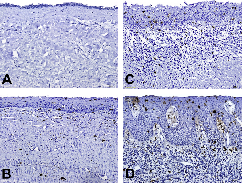
BACKGROUND/PURPOSE: Langerhans cells (LCs) are antigen-presenting cells. This study assessed the LC counts in 80 dentigerous cysts (DCs).

MATERIALS AND METHODS

The S100-positive LC numbers in the lining epithelia and subepithelial connective tissues were counted at 80 DC sites without inflammation, 33 DC sites with mild/moderate inflammation, and 9 DC sites with severe inflammation from 80 DC specimens.

RESULTS

The mean S100-positive LC counts in the lining epithelia and subepithelial connective tissues increased significantly from no inflammation (0.6 ± 0.6 and 0.7 ± 0.6 cell/high-power field or HPF, respectively) through mild/moderate inflammation (8.1 ± 2.0 and 4.5 ± 2.3 cells/HPF, respectively) to severe inflammation DC sites (21.0 ± 7.0 and 11.1 ± 6.5 cells/HPF, respectively; -value < 0.001). DC sites with inflammation had thicker lining epithelia than those without inflammation. Moreover, the mean LC counts in the lining epithelia and subepithelial connective tissues of DCs were significantly higher in the thicker lining epithelium (>50 μm) group (8.6 ± 7.1 and 4.8 ± 4.5 cells/HPF, respectively) than in the thinner lining epithelium (≦50 μm) group (0.6 ± 0.6 and 0.6 ± 0.6 cells/HPF, respectively; both -values < 0.001).

CONCLUSION

A significant association of high-grade inflammation and thick lining epithelium with the increased LC number in DCs is found. Very few LCs in the lining epithelia of DCs without inflammation indicate the reduced immunosurveillance ability against DC lining epithelial cells in DC patients. It needs further studies to confirm the role of reduced immunosurveillance in the enlargement of the DC.

摘要

背景/目的:朗格汉斯细胞(LCs)是抗原呈递细胞。本研究评估了80个含牙囊肿(DCs)中的LC计数。

材料与方法

从80个DC标本中,对80个无炎症的DC部位、33个有轻度/中度炎症的DC部位和9个有重度炎症的DC部位的衬里上皮和上皮下结缔组织中S100阳性LC数量进行计数。

结果

衬里上皮和上皮下结缔组织中S100阳性LC的平均计数从无炎症(分别为0.6±0.6和0.7±0.6个细胞/高倍视野或HPF)经轻度/中度炎症(分别为8.1±2.0和4.5±2.3个细胞/HPF)到重度炎症的DC部位(分别为21.0±7.0和11.1±6.5个细胞/HPF)显著增加(P值<0.001)。有炎症的DC部位的衬里上皮比无炎症的更厚。此外,DCs衬里上皮和上皮下结缔组织中LC的平均计数在较厚衬里上皮(>50μm)组(分别为8.6±7.1和4.8±4.5个细胞/HPF)显著高于较薄衬里上皮(≤50μm)组(分别为0.6±0.6和0.6±0.6个细胞/HPF;两个P值均<0.001)。

结论

发现DCs中高级别炎症和厚衬里上皮与LC数量增加显著相关。无炎症的DC衬里上皮中LC极少表明DC患者对DC衬里上皮细胞的免疫监视能力降低。需要进一步研究来证实免疫监视降低在DC扩大中的作用。

https://cdn.ncbi.nlm.nih.gov/pmc/blobs/6bfc/6395349/b68ca910e04c/gr1.jpg

相似文献

1
S100 protein-positive Langerhans cells in 80 dentigerous cysts.80例含牙囊肿中的S100蛋白阳性朗格汉斯细胞
J Dent Sci. 2017 Dec;12(4):405-412. doi: 10.1016/j.jds.2017.08.001. Epub 2017 Sep 25.
2
Significant association of high-grade inflammation and thick lining epithelium with the increased number of Langerhans cells in dentigerous cysts.高等级炎症和厚衬里上皮与含牙囊肿中朗格汉斯细胞数量增加有显著关联。
J Formos Med Assoc. 2017 Nov;116(11):837-843. doi: 10.1016/j.jfma.2017.06.011. Epub 2017 Jul 24.
3
Significant association of inflammation grade with the number of Langerhans cells in odontogenic keratocysts.炎症程度与牙源性角化囊肿中朗格汉斯细胞数量存在显著相关性。
J Formos Med Assoc. 2017 Oct;116(10):798-805. doi: 10.1016/j.jfma.2017.04.002. Epub 2017 Jul 26.
4
Langerhans cells in 60 odontogenic keratocysts.60例牙源性角化囊肿中的朗格汉斯细胞。
J Dent Sci. 2017 Sep;12(3):283-290. doi: 10.1016/j.jds.2017.04.001. Epub 2017 May 9.
5
Langerhans cell counts in oral epithelial dysplasia and their correlation to clinicopathological parameters.口腔上皮发育异常中的朗格汉斯细胞计数及其与临床病理参数的相关性。
J Formos Med Assoc. 2017 Jun;116(6):457-463. doi: 10.1016/j.jfma.2017.02.006. Epub 2017 Mar 11.
6
Immunohistochemical expression of SHH, PTC, SMO and GLI1 in glandular odontogenic cysts and dentigerous cysts.在腺牙源性囊肿和含牙囊肿中 SHH、PTC、SMO 和 GLI1 的免疫组织化学表达。
Oral Dis. 2010 Nov;16(8):818-22. doi: 10.1111/j.1601-0825.2010.01697.x.
7
A molecular insight into the role of inflammation in the behavior and pathogenesis of odontogenic cysts.对炎症在牙源性囊肿的行为和发病机制中的作用的分子见解。
Ann Med Health Sci Res. 2013 Oct;3(4):523-8. doi: 10.4103/2141-9248.122072.
8
Tumor-infiltrating macrophages and dendritic cells in human colorectal cancer: relation to local regulatory T cells, systemic T-cell response against tumor-associated antigens and survival.人类结直肠癌中的肿瘤浸润巨噬细胞和树突状细胞:与局部调节性T细胞、针对肿瘤相关抗原的全身T细胞反应及生存的关系
J Transl Med. 2007 Nov 29;5:62. doi: 10.1186/1479-5876-5-62.
9
High expression of osteopontin and CD44v6 in odontogenic keratocysts.骨桥蛋白和CD44v6在牙源性角化囊肿中的高表达。
J Formos Med Assoc. 2009 Apr;108(4):286-92. doi: 10.1016/S0929-6646(09)60068-1.
10
Convolutions of dentigerous cyst: An institutional experience.含牙囊肿的影像学表现:一项机构经验。
J Exp Ther Oncol. 2019 Jan;13(1):65-70.

引用本文的文献

1
The Tilapia Cyst Tissue Enclosing the Proliferating Parasite Exhibits Cornified Structure and Immune Barrier Function.罗非鱼囊包增殖寄生虫的囊组织具有角质化结构和免疫屏障功能。
Int J Mol Sci. 2024 May 23;25(11):5683. doi: 10.3390/ijms25115683.
2
Oral giant cell fibroma: A report of three cases.口腔巨细胞纤维瘤:三例报告
J Dent Sci. 2021 Jan;16(1):552-554. doi: 10.1016/j.jds.2020.09.001. Epub 2020 Sep 14.
3
Marking vertebrates langerhans cells, from fish to mammals.标记鱼类到哺乳动物的朗格汉斯细胞。

本文引用的文献

1
Langerhans cells in 60 odontogenic keratocysts.60例牙源性角化囊肿中的朗格汉斯细胞。
J Dent Sci. 2017 Sep;12(3):283-290. doi: 10.1016/j.jds.2017.04.001. Epub 2017 May 9.
2
Langerhans cells in plexiform ameloblastoma.丛状成釉细胞瘤中的朗格汉斯细胞。
J Dent Sci. 2017 Jun;12(2):195-197. doi: 10.1016/j.jds.2017.02.001. Epub 2017 Mar 14.
3
Non-calcifying and Langerhans cell-rich variant of calcifying epithelial odontogenic tumor.牙源性钙化上皮瘤的非钙化且富含朗格汉斯细胞变体
Acta Histochem. 2020 Oct;122(7):151622. doi: 10.1016/j.acthis.2020.151622. Epub 2020 Sep 9.
4
Cicatricial pemphigoid presenting as desquamative gingivitis.表现为剥脱性龈炎的瘢痕性类天疱疮。
J Dent Sci. 2020 Mar;15(1):110-111. doi: 10.1016/j.jds.2019.05.004. Epub 2019 Dec 23.
5
Stafne bone defect of the molar region of the mandible.下颌磨牙区斯丹福骨缺损
J Dent Sci. 2019 Dec;14(4):378-382. doi: 10.1016/j.jds.2019.05.002. Epub 2019 Jun 13.
6
Hindrance of tooth eruption and orthodontic tooth movement by focal idiopathic osteosclerosis in the mandible.下颌骨局灶性特发性骨硬化对牙齿萌出及正畸牙齿移动的阻碍
J Dent Sci. 2019 Sep;14(3):332-334. doi: 10.1016/j.jds.2019.02.001. Epub 2019 Mar 20.
7
Huge lipoma in the left submandibular region.左下颌下区巨大脂肪瘤。
J Dent Sci. 2019 Sep;14(3):330-331. doi: 10.1016/j.jds.2019.01.004. Epub 2019 Mar 22.
8
Lymphoepithelial cyst of the tongue.舌淋巴上皮囊肿
J Dent Sci. 2019 Jun;14(2):209-210. doi: 10.1016/j.jds.2019.02.003. Epub 2019 Mar 20.
J Dent Sci. 2016 Jun;11(2):117-122. doi: 10.1016/j.jds.2016.02.001. Epub 2016 Mar 28.
4
Significant association of inflammation grade with the number of Langerhans cells in odontogenic keratocysts.炎症程度与牙源性角化囊肿中朗格汉斯细胞数量存在显著相关性。
J Formos Med Assoc. 2017 Oct;116(10):798-805. doi: 10.1016/j.jfma.2017.04.002. Epub 2017 Jul 26.
5
Significant association of high-grade inflammation and thick lining epithelium with the increased number of Langerhans cells in dentigerous cysts.高等级炎症和厚衬里上皮与含牙囊肿中朗格汉斯细胞数量增加有显著关联。
J Formos Med Assoc. 2017 Nov;116(11):837-843. doi: 10.1016/j.jfma.2017.06.011. Epub 2017 Jul 24.
6
Langerhans cell counts in oral epithelial dysplasia and their correlation to clinicopathological parameters.口腔上皮发育异常中的朗格汉斯细胞计数及其与临床病理参数的相关性。
J Formos Med Assoc. 2017 Jun;116(6):457-463. doi: 10.1016/j.jfma.2017.02.006. Epub 2017 Mar 11.
7
Langerhans cells in granular cell ameloblastoma.颗粒细胞成釉细胞瘤中的朗格汉斯细胞。
J Formos Med Assoc. 2017 Aug;116(8):642-644. doi: 10.1016/j.jfma.2017.02.007. Epub 2017 Mar 9.
8
Langerhans cells in a dermoid cyst epithelium lining.皮样囊肿上皮衬里中的朗格汉斯细胞。
J Formos Med Assoc. 2016 Jan;115(1):57-8. doi: 10.1016/j.jfma.2015.08.014. Epub 2015 Oct 4.
9
Noncalcifying variant of calcifying epithelial odontogenic tumor with Langerhans cells.伴有朗格汉斯细胞的钙化上皮性牙源性肿瘤的非钙化变体
J Formos Med Assoc. 2015 Aug;114(8):781-2. doi: 10.1016/j.jfma.2015.04.004. Epub 2015 May 18.
10
Langerhans cells in keratoacanthoma.角化棘皮瘤中的朗格汉斯细胞。
J Formos Med Assoc. 2015 May;114(5):475-6. doi: 10.1016/j.jfma.2014.12.008. Epub 2015 Feb 7.