Foley Elaine, Cross J Helen, Thai Ngoc J, Walsh A Richard, Bill Peter, Furlong Paul, Wood Amanda G, Cerquiglini Antonella, Seri Stefano
Aston Brain Centre, School of Life and Health Sciences, Aston University, Birmingham, UK.
Developmental Neurosciences Programme, Institute of Child Health, University College London, London, UK.
Brain Topogr. 2019 May;32(3):492-503. doi: 10.1007/s10548-019-00703-1. Epub 2019 Mar 20.
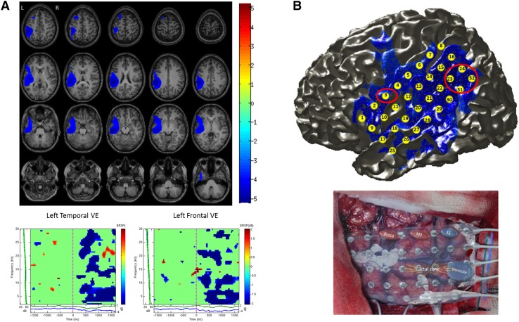
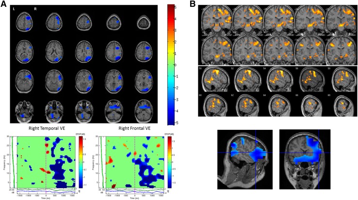
Establishing language dominance is an important step in the presurgical evaluation of patients with refractory epilepsy. In the absence of a universally accepted gold-standard non-invasive method to determine language dominance in the preoperative assessment, a range of tools and methodologies have recently received attention. When applied to pediatric age, many of the proposed methods, such as functional magnetic resonance imaging (fMRI), may present some challenges due to the time-varying effects of epileptogenic lesions and of on-going seizures on maturational phenomena. Magnetoencephalography (MEG) has the advantage of being insensitive to the distortive effects of anatomical lesions on brain microvasculature and to differences in the metabolism or vascularization of the developing brain and also provides a less intimidating recording environment for younger children. In this study we investigated the reliability of lateralized synchronous cortical activation during a verb generation task in a group of 28 children (10 males and 18 females, mean age 12 years) with refractory epilepsy who were evaluated for epilepsy surgery. The verb generation task was associated with significant decreases in beta oscillatory power (13-30 Hz) in frontal and temporal lobes. The MEG data were compared with other available presurgical non-invasive data including cortical stimulation, neuropsychological and fMRI data on language lateralization where available. We found that the lateralization of MEG beta power reduction was concordant with language dominance determined by one or more different assessment methods (i.e. cortical stimulation mapping, neuropsychological, fMRI or post-operative data) in 89% of patients. Our data suggest that qualitative hemispheric differences in task-related changes of spectral power could offer a promising insight into the contribution of dominant and non-dominant hemispheres in language processing and may help to characterize the specialization and lateralization of language processes in children.
确定语言优势半球是难治性癫痫患者术前评估的重要一步。在术前评估中,目前尚无一种被普遍接受的用于确定语言优势半球的金标准非侵入性方法,一系列工具和方法最近受到了关注。当应用于儿童时,许多所提出的方法,如功能磁共振成像(fMRI),可能会因致痫性病变和正在发作的癫痫对成熟现象的时变影响而面临一些挑战。脑磁图(MEG)的优势在于对解剖病变对脑微血管的扭曲作用以及发育中大脑代谢或血管化差异不敏感,并且为年幼儿童提供了一个不那么令人生畏的记录环境。在本研究中,我们调查了一组28名接受癫痫手术评估的难治性癫痫儿童(10名男性和18名女性,平均年龄12岁)在动词生成任务期间侧化同步皮质激活的可靠性。动词生成任务与额叶和颞叶β振荡功率(13 - 30Hz)的显著降低相关。将MEG数据与其他可用的术前非侵入性数据进行比较,包括皮质刺激、神经心理学以及在可用时关于语言侧化的fMRI数据。我们发现,在89%的患者中,MEGβ功率降低的侧化与通过一种或多种不同评估方法(即皮质刺激映射、神经心理学、fMRI或术后数据)确定的语言优势半球一致。我们的数据表明,任务相关频谱功率变化中的定性半球差异可能为优势半球和非优势半球在语言处理中的作用提供有前景的见解,并可能有助于刻画儿童语言过程的专业化和侧化特征。