Experimental Molecular Biophysics, Department of Physics, Freie Universität Berlin, Berlin, Germany.
Division of Biology and Chemistry-Laboratory for Biomolecular Research, Paul Scherrer Institute, Villigen, Switzerland; Department of Biology, ETH Zürich, Zürich, Switzerland.
Biophys J. 2019 Apr 2;116(7):1248-1258. doi: 10.1016/j.bpj.2019.02.025. Epub 2019 Mar 5.
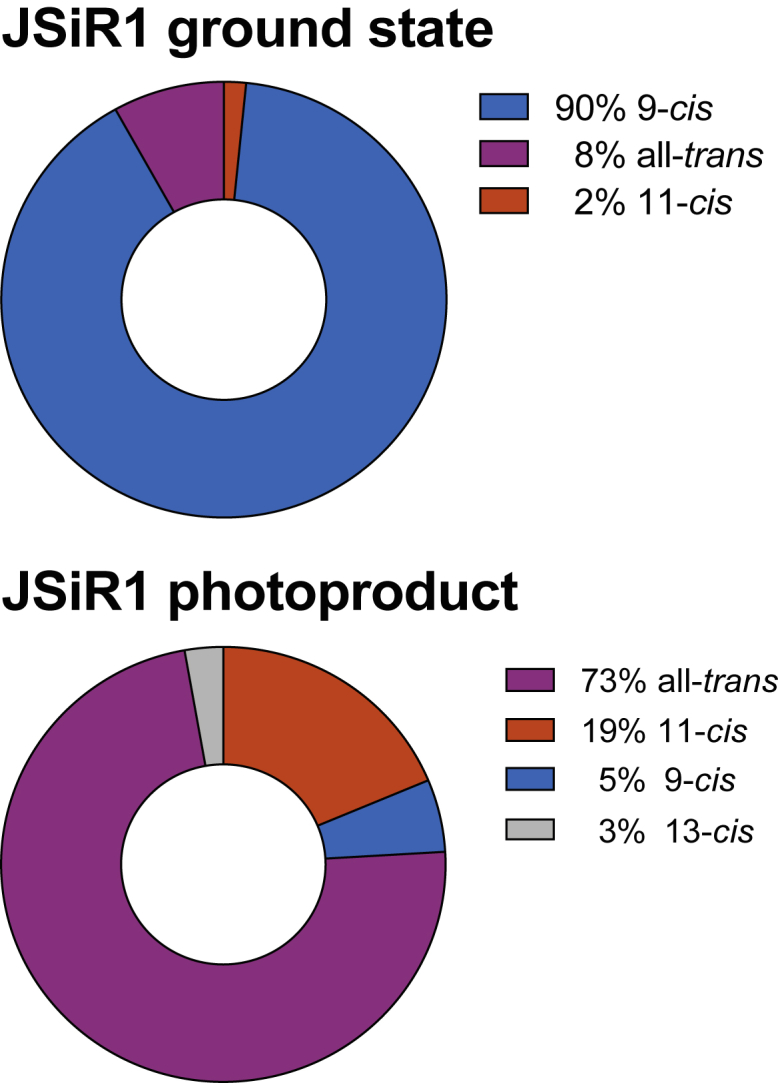
Bistable opsins are photopigments expressed in both invertebrates and vertebrates. These light-sensitive G-protein-coupled receptors undergo a reversible reaction upon illumination. A first photon initiates the cis to trans isomerization of the retinal chromophore-attached to the protein through a protonated Schiff base-and a series of transition states that eventually results in the formation of the thermally stable and active Meta state. Excitation by a second photon reverts this process to recover the original ground state. On the other hand, monostable opsins (e.g., bovine rhodopsin) lose their chromophore during the decay of the Meta II state (i.e., they bleach). Spectroscopic studies on the molecular details of the two-photon cycle in bistable opsins are limited. Here, we describe the successful expression and purification of recombinant rhodopsin-1 from the jumping spider Hasarius adansoni (JSR1). In its natural configuration, spectroscopic characterization of JSR1 is hampered by the similar absorption spectra in the visible spectrum of the inactive and active states. We solved this issue by separating their absorption spectra by replacing the endogenous 11-cis retinal chromophore with the blue-shifted 9-cis JSiR1. With this system, we used time-resolved ultraviolet-visible spectroscopy after pulsed laser excitation to obtain kinetic details of the rise and decay of the photocycle intermediates. We also used resonance Raman spectroscopy to elucidate structural changes of the retinal chromophore upon illumination. Our data clearly indicate that the protonated Schiff base is stable throughout the entire photoreaction. We additionally show that the accompanying conformational changes in the protein are different from those of monostable rhodopsin, as recorded by light-induced FTIR difference spectroscopy. Thus, we envisage JSR1 as becoming a model system for future studies on the reaction mechanisms of bistable opsins, e.g., by time-resolved x-ray crystallography.
双稳态视蛋白存在于无脊椎动物和脊椎动物中。这些对光敏感的 G 蛋白偶联受体在光照下会发生可逆反应。第一个光子通过质子化的 Schiff 碱引发与蛋白结合的视黄醛发色团的顺式到反式异构化,以及一系列过渡态,最终导致形成热稳定和活跃的 Meta 状态。第二个光子的激发使这个过程反转,恢复原来的基态。另一方面,单稳态视蛋白(例如牛视紫红质)在 Meta II 状态衰减过程中失去其发色团(即漂白)。关于双稳态视蛋白中双光子循环的分子细节的光谱研究受到限制。在这里,我们描述了来自跳蛛 Hasarius adansoni(JSR1)的重组视紫红质-1 的成功表达和纯化。在其自然构象中,JSR1 的光谱特征受到非活性和活性状态下相似可见光谱吸收谱的阻碍。我们通过用蓝移的 9-cis JSiR1 取代内源性 11-cis 视黄醛来分离它们的吸收光谱,从而解决了这个问题。使用该系统,我们在脉冲激光激发后使用时间分辨紫外可见光谱获得光循环中间体的上升和衰减的动力学细节。我们还使用共振拉曼光谱阐明了光照下视黄醛发色团的结构变化。我们的数据清楚地表明,质子化的 Schiff 碱在整个光反应过程中是稳定的。我们还表明,与单稳态视紫红质不同,蛋白伴随的构象变化,如用光诱导的 FTIR 差光谱记录的那样。因此,我们设想 JSR1 成为未来研究双稳态视蛋白反应机制的模型系统,例如通过时间分辨 X 射线晶体学。