Department of Anesthesiology, Tianjin Medical University Cancer Institute and Hospital, National Clinical Research Center of Cancer, Tianjin 300060, China.
Key Laboratory of Cancer Prevention and Therapy, Tianjin 300060, China.
Oxid Med Cell Longev. 2019 Feb 14;2019:9078209. doi: 10.1155/2019/9078209. eCollection 2019.
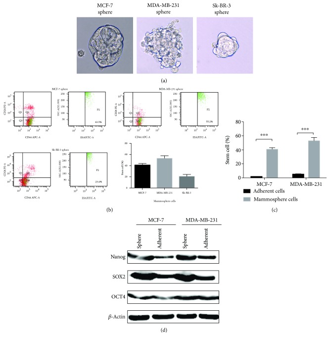
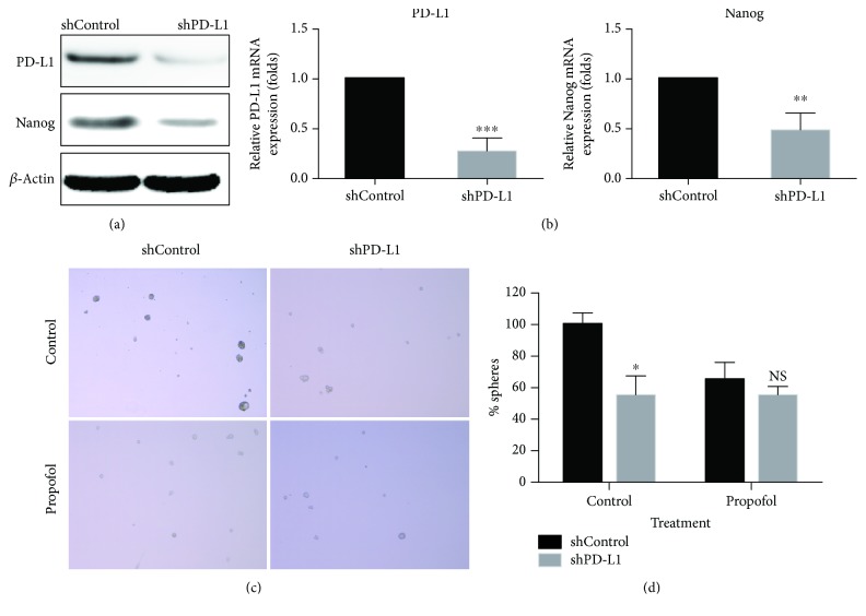
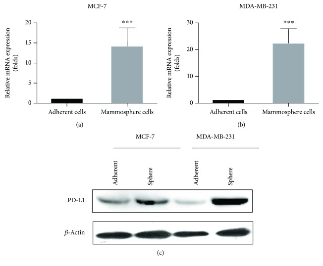
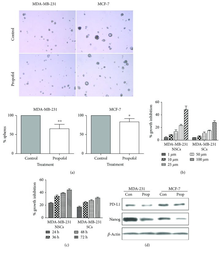
Several researches revealed that propofol, a hypnotic intravenous anesthesia agent, could inhibit the cancer cell proliferation and tumor formation, which might affect cancer recurrence or metastasis and impact patients' prognosis. Cancer stem cells (CSCs) comprised a tiny fraction of tumor bulk and played a vital role in cancer recurrence and eventual mortality. This study investigates the effect of propofol on breast cancer stem cells (BCSCs) and the underlying molecular mechanisms. Tumor formation of CSCs was measured by mammosphere culture. Cultured BCSCs were exposed to different concentrations and durations of propofol. Cell proliferation and self-renewal capacity were determined by MTT assays. Expressions of PD-L1 and Nanog were measured using western blotting and real-time PCR. We knocked down the PD-L1 expression in MDA-MB-231 cells by lentivirus-mediated RNAi technique, and the mammosphere-forming ability of shControl and shPD-L1 under propofol treatment was examined. Mammosphere culture could enrich BCSCs. Compared with control, cells exposed to propofol for 24 h induced a larger number of mammosphere cells ( = 0.0072). Levels of PD-L1 and Nanog were downregulated by propofol. Compared with shControl stem cells, there was no significant difference in the inhibitory effect of propofol on the mammosphere-forming ability of shPD-L1 stem cells which indicated that the inhibition of propofol could disappear in PD-L1 knockdown breast stem cells. Propofol could reduce the mammosphere-forming ability of BCSCs . Mechanism experiments indicated that the inhibition of propofol in mammosphere formation of BCSCs might be mediated through PD-L1, which was important to maintain Nanog.
几项研究表明,作为一种静脉麻醉剂的异丙酚能够抑制癌细胞增殖和肿瘤形成,这可能会影响癌症的复发或转移,并影响患者的预后。癌症干细胞(CSCs)构成肿瘤块的一小部分,在癌症复发和最终死亡中起着至关重要的作用。本研究探讨了异丙酚对乳腺癌干细胞(BCSCs)的影响及其潜在的分子机制。通过乳腺球体培养来测量 CSCs 的肿瘤形成。将培养的 BCSCs 暴露于不同浓度和时间的异丙酚中。通过 MTT 测定法来测定细胞增殖和自我更新能力。使用 Western blot 和实时 PCR 来测量 PD-L1 和 Nanog 的表达。我们通过慢病毒介导的 RNAi 技术敲低 MDA-MB-231 细胞中的 PD-L1 表达,并在异丙酚处理下检测 shControl 和 shPD-L1 的乳腺球体形成能力。乳腺球体培养可以富集 BCSCs。与对照组相比,暴露于异丙酚 24 小时的细胞诱导出更多的乳腺球体细胞(=0.0072)。PD-L1 和 Nanog 的水平被异丙酚下调。与 shControl 干细胞相比,shPD-L1 干细胞中异丙酚对乳腺球体形成能力的抑制作用没有显著差异,这表明 PD-L1 敲低的乳腺癌干细胞中异丙酚的抑制作用消失。异丙酚可以降低 BCSCs 的乳腺球体形成能力。机制实验表明,异丙酚抑制 BCSCs 乳腺球体形成可能是通过 PD-L1 介导的,PD-L1 对维持 Nanog 很重要。