Second Clinical Medical College of Zhejiang Chinese Medical University, Hangzhou 310053, China.
College of Life Sciences of Zhejiang Chinese Medical University, Hangzhou 310053, China.
Biomed Res Int. 2019 Feb 17;2019:5094842. doi: 10.1155/2019/5094842. eCollection 2019.
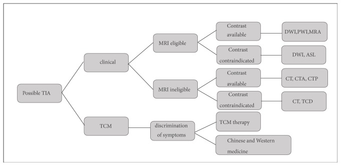
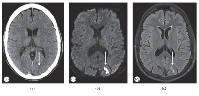
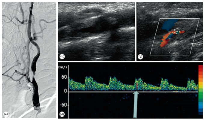
Neuroimaging plays a pivotal role in Transient Ischemic Attack (TIA). Generally, clinicians focus on the specific changes in morphology and function, but the diagnosis of TIA often depends on imaging evidence. Whereas Traditional Chinese Medicine (TCM) is concerned with the performance of clinical symptoms, they began to use imaging methods to diagnose TIA. CT and MRI are the recommended modality to diagnose TIA and image ischemic lesions. In addition, Transcranial Doppler sonography (TCD) and Digital Subtraction Angiography (DSA) are two acceptable alternatives for diagnosing TIA patients. This article elaborates the update of imaging modalities in clinic and the development of imaging modalities in TCM. Besides, multiple joint imaging technologies also will be evaluated whether enhanced diagnostic yields availably.
神经影像学在短暂性脑缺血发作(TIA)中起着关键作用。一般来说,临床医生关注形态和功能的特定变化,但 TIA 的诊断通常依赖于影像学证据。而中医(TCM)则关注临床症状的表现,他们开始使用影像学方法来诊断 TIA。CT 和 MRI 是诊断 TIA 和显示缺血性病变的推荐方法。此外,经颅多普勒超声(TCD)和数字减影血管造影(DSA)是诊断 TIA 患者的两种可接受的替代方法。本文阐述了临床影像学方法的更新和 TCM 影像学方法的发展。此外,还将评估多种联合成像技术是否能有效地提高诊断效果。