• 文献检索
  • 文档翻译
  • 深度研究
  • 学术资讯
  • Suppr Zotero 插件Zotero 插件
  • 邀请有礼
  • 套餐&价格
  • 历史记录
应用&插件
Suppr Zotero 插件Zotero 插件浏览器插件Mac 客户端Windows 客户端微信小程序
定价
高级版会员购买积分包购买API积分包
服务
文献检索文档翻译深度研究API 文档MCP 服务
关于我们
关于 Suppr公司介绍联系我们用户协议隐私条款
关注我们

Suppr 超能文献

核心技术专利:CN118964589B侵权必究
粤ICP备2023148730 号-1Suppr @ 2026

文献检索

告别复杂PubMed语法,用中文像聊天一样搜索,搜遍4000万医学文献。AI智能推荐,让科研检索更轻松。

立即免费搜索

文件翻译

保留排版,准确专业,支持PDF/Word/PPT等文件格式,支持 12+语言互译。

免费翻译文档

深度研究

AI帮你快速写综述,25分钟生成高质量综述,智能提取关键信息,辅助科研写作。

立即免费体验

基于撕裂间隙的退行性内侧半月板后根撕裂的新型关节镜分类

A Novel Arthroscopic Classification of Degenerative Medial Meniscus Posterior Root Tears Based on the Tear Gap.

作者信息

Kim Jae-Young, Bin Seong-Il, Kim Jong-Min, Lee Bum-Sik, Oh Sung-Mok, Cho Won-Joon

机构信息

Department of Orthopedic Surgery, Asan Medical Center, University of Ulsan College of Medicine, Seoul, Republic of Korea.

出版信息

Orthop J Sports Med. 2019 Mar 18;7(3):2325967119827945. doi: 10.1177/2325967119827945. eCollection 2019 Mar.

DOI:10.1177/2325967119827945
PMID:30911565
原文链接:https://pmc.ncbi.nlm.nih.gov/articles/PMC6423685/
Abstract

BACKGROUND

Degenerative medial meniscus posterior root tears (MMPRTs) are reportedly associated with medial compartment osteoarthritis and meniscal extrusion with a displaced gap from the root insertion. However, degenerative MMPRTs have not yet been clearly classified according to arthroscopic findings.

PURPOSE

To classify degenerative MMPRTs according to the tear gap and to investigate how the classification could reflect the joint condition properly.

STUDY DESIGN

Cohort study; Level of evidence, 3.

METHODS

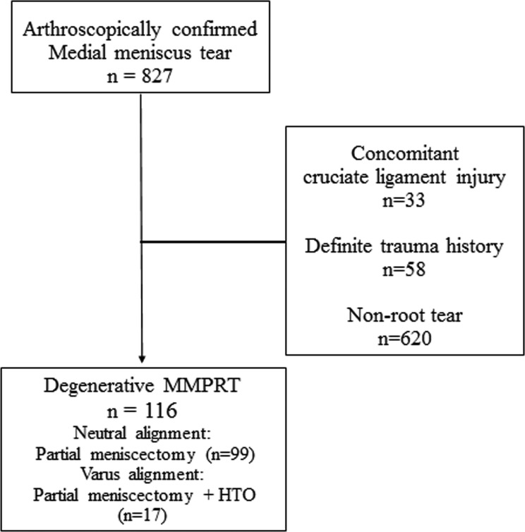
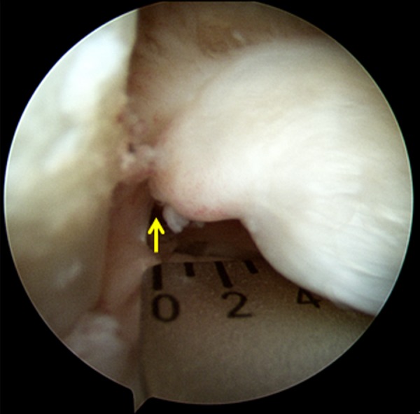
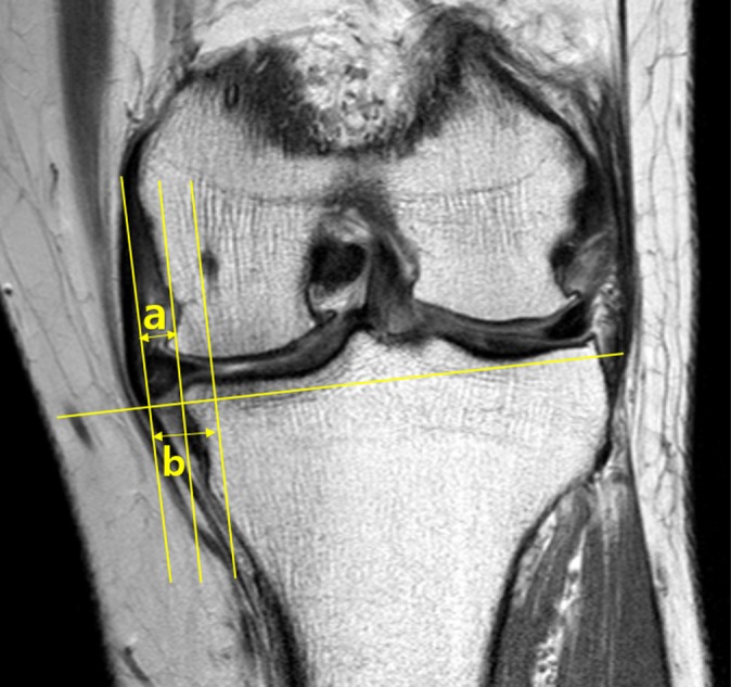
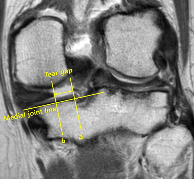
Patients who underwent arthroscopic surgery, performed by a single orthopaedic surgeon, for degenerative MMPRTs between August 2006 and February 2017 were included. MMPRTs were classified according to tear patterns observed during arthroscopic surgery (type 1, incomplete root tear; types 2-5, complete root tears), with each type further divided by the size of the tear gap, defined as the degree of tear displacement from the root (type 2, no gap or overlapped; type 3, gap of 1-3 mm; type 4, gap of 4-6 mm; type 5, gap of ≥7 mm). We compared preoperative factors, including the Kellgren-Lawrence (K-L) grade, absolute extrusion, relative percentage of extrusion (RPE), tear gap on magnetic resonance imaging (MRI), and mechanical alignment, as well as intraoperative factors, including chondral wear at surgery, between each MMPRT type.

RESULTS

A total of 116 root tears were categorized according to this classification: type 1, 16.4% (19 knees); type 2, 9.5% (11 knees); type 3, 40.5% (47 knees); type 4, 25.0% (29 knees); and type 5, 8.6% (10 knees). Chondral wear of the medial femoral condyle (MFC) ( = .001), K-L grade ( = .001), meniscal extrusion ( = .001), and tear gap on MRI ( = .001) showed a tendency to increase with a higher tear type. Chondral wear (ρ for MFC = 0.388; ρ for MTP = 0.311), K-L grade (ρ = 0.390), and meniscal extrusion (ρ for absolute extrusion = 0.500; ρ for RPE = 0.451) showed a moderate correlation with tear type, whereas tear gap on MRI (ρ = 0.907) showed a strong correlation with tear type.

CONCLUSION

Our study introduces a new classification based on the tear gap that can concisely describe a degenerative MMPRT. The classification system demonstrated that a higher tear type (increasing displacement of the tear gap in arthroscopic surgery) is associated with higher meniscal extrusion, severe chondral wear, and greater severity of arthritis.

摘要

背景

据报道,退行性内侧半月板后根撕裂(MMPRTs)与内侧间室骨关节炎和半月板挤出有关,且根部插入处存在移位间隙。然而,根据关节镜检查结果,退行性MMPRTs尚未得到明确分类。

目的

根据撕裂间隙对退行性MMPRTs进行分类,并研究该分类如何恰当地反映关节状况。

研究设计

队列研究;证据等级,3级。

方法

纳入2006年8月至2017年2月间由一名骨科医生进行关节镜手术治疗退行性MMPRTs的患者。根据关节镜手术中观察到的撕裂模式对MMPRTs进行分类(1型,不完全根部撕裂;2 - 5型,完全根部撕裂),每种类型再根据撕裂间隙大小进一步划分,撕裂间隙定义为从根部的撕裂移位程度(2型,无间隙或重叠;3型,间隙为1 - 3毫米;4型,间隙为4 - 6毫米;5型,间隙≥7毫米)。我们比较了术前因素,包括凯尔格伦 - 劳伦斯(K - L)分级、绝对挤出量、相对挤出百分比(RPE)、磁共振成像(MRI)上的撕裂间隙以及机械对线情况,以及术中因素,包括手术时软骨磨损情况,比较各MMPRT类型之间的差异。

结果

根据该分类共对116例根部撕裂进行了分类:1型,16.4%(19膝);2型,9.5%(11膝);3型,40.5%(47膝);4型,25.0%(29膝);5型,8.6%(10膝)。股骨内侧髁(MFC)的软骨磨损(P = 0.001)、K - L分级(P = 0.001)、半月板挤出(P = 0.001)以及MRI上的撕裂间隙(P = 0.001)均呈现出随着撕裂类型增高而增加的趋势。软骨磨损(MFC的ρ = 0.388;胫骨平台内侧的ρ = 0.311)、K - L分级(ρ = 0.390)以及半月板挤出(绝对挤出量的ρ = 0.500;RPE的ρ = 0.451)与撕裂类型呈中度相关,而MRI上的撕裂间隙(ρ = 0.907)与撕裂类型呈强相关。

结论

我们的研究引入了一种基于撕裂间隙的新分类方法,该方法能够简洁地描述退行性MMPRTs。该分类系统表明,更高的撕裂类型(关节镜手术中撕裂间隙移位增加)与更高的半月板挤出、严重的软骨磨损以及更严重的关节炎相关。

https://cdn.ncbi.nlm.nih.gov/pmc/blobs/56ea/6423685/129fcc025311/10.1177_2325967119827945-fig5.jpg
https://cdn.ncbi.nlm.nih.gov/pmc/blobs/56ea/6423685/80e7035bc9b1/10.1177_2325967119827945-fig1.jpg
https://cdn.ncbi.nlm.nih.gov/pmc/blobs/56ea/6423685/9a3954bd2e51/10.1177_2325967119827945-fig2.jpg
https://cdn.ncbi.nlm.nih.gov/pmc/blobs/56ea/6423685/2e2d7c8865e2/10.1177_2325967119827945-fig3.jpg
https://cdn.ncbi.nlm.nih.gov/pmc/blobs/56ea/6423685/98623e4b6b9e/10.1177_2325967119827945-fig4.jpg
https://cdn.ncbi.nlm.nih.gov/pmc/blobs/56ea/6423685/129fcc025311/10.1177_2325967119827945-fig5.jpg

相似文献

1
A Novel Arthroscopic Classification of Degenerative Medial Meniscus Posterior Root Tears Based on the Tear Gap.基于撕裂间隙的退行性内侧半月板后根撕裂的新型关节镜分类
Orthop J Sports Med. 2019 Mar 18;7(3):2325967119827945. doi: 10.1177/2325967119827945. eCollection 2019 Mar.
2
Tear gap and severity of osteoarthritis are associated with meniscal extrusion in degenerative medial meniscus posterior root tears.半月板后根撕裂中,撕裂间隙和骨关节炎严重程度与半月板外突有关。
Orthop Traumatol Surg Res. 2019 Nov;105(7):1395-1399. doi: 10.1016/j.otsr.2019.09.015. Epub 2019 Sep 28.
3
A new arthroscopic classification of degenerative medial meniscus root tear that correlates with meniscus extrusion on magnetic resonance imaging.一种与磁共振成像上半月板挤出相关的退行性内侧半月板根部撕裂的新关节镜分类。
Knee. 2016 Mar;23(2):246-50. doi: 10.1016/j.knee.2015.07.003. Epub 2015 Aug 1.
4
Investigating the Chronology of Meniscus Root Tears: Do Medial Meniscus Posterior Root Tears Cause Extrusion or the Other Way Around?探究半月板根部撕裂的时间顺序:内侧半月板后根撕裂会导致挤压,还是反之亦然?
Orthop J Sports Med. 2020 Nov 4;8(11):2325967120961368. doi: 10.1177/2325967120961368. eCollection 2020 Nov.
5
Predictors of degenerative medial meniscus extrusion: radial component and knee osteoarthritis.预测内侧半月板退行性外突的因素:放射学成分和膝关节骨关节炎。
Knee Surg Sports Traumatol Arthrosc. 2011 Feb;19(2):222-9. doi: 10.1007/s00167-010-1274-2. Epub 2010 Oct 2.
6
Correlation between meniscal extrusion and symptom duration, alignment, and arthritic changes in medial meniscus posterior root tear: research article.内侧半月板后根撕裂中半月板挤出与症状持续时间、对线及关节炎改变之间的相关性:研究文章
Knee Surg Relat Res. 2020 Jan 1;32(1):2. doi: 10.1186/s43019-019-0019-x.
7
Magnetic resonance imaging evidence of meniscal extrusion in medial meniscus posterior root tear.内侧半月板后根撕裂的半月板挤压的磁共振成像证据。
Arthroscopy. 2010 Dec;26(12):1602-6. doi: 10.1016/j.arthro.2010.05.004.
8
Medial knee osteoarthritis precedes medial meniscal posterior root tear with an event of painful popping.内侧膝关节骨关节炎先于内侧半月板后根撕裂出现疼痛性弹响。
Orthop Traumatol Surg Res. 2018 Nov;104(7):1009-1015. doi: 10.1016/j.otsr.2018.07.010. Epub 2018 Aug 11.
9
Medial meniscus extrusion and stage are related to the size of spontaneous osteonecrosis of the knee in patients who underwent high tibial osteotomy.内侧半月板突出和分期与接受胫骨高位截骨术的膝关节自发性骨坏死患者的骨坏死大小有关。
Knee. 2022 Jun;36:72-79. doi: 10.1016/j.knee.2022.04.011. Epub 2022 May 6.
10
Characteristic location and rapid progression of medial femoral condylar chondral lesions accompanying medial meniscus posterior root tear.股骨内侧髁软骨损伤伴随内侧半月板后根撕裂的特征性位置及快速进展
Knee. 2019 Jun;26(3):673-678. doi: 10.1016/j.knee.2019.03.003. Epub 2019 Mar 21.

引用本文的文献

1
The preoperative flexion tear gap affects postoperative meniscus stability after pullout repair for medial meniscus posterior root tear.术前屈曲时的撕裂间隙会影响内侧半月板后根撕裂拔出修复术后半月板的稳定性。
Knee Surg Relat Res. 2025 Apr 3;37(1):16. doi: 10.1186/s43019-025-00264-7.
2
Progression of patellofemoral joint cartilage degeneration within 1 year after medial meniscus posterior root repair: A retrospective study.内侧半月板后根修复术后1年内髌股关节软骨退变的进展:一项回顾性研究。
J Exp Orthop. 2025 Apr 1;12(2):e70139. doi: 10.1002/jeo2.70139. eCollection 2025 Apr.
3
Fast rehabilitation does not worsen clinical, radiological, and arthroscopic outcomes after medial meniscus posterior root repair: A retrospective comparative study.

本文引用的文献

1
Medial meniscus extrusion correlates with disease duration of the sudden symptomatic medial meniscus posterior root tear.内侧半月板后角撕裂突发症状与内侧半月板挤压的病程相关。
Orthop Traumatol Surg Res. 2017 Dec;103(8):1179-1182. doi: 10.1016/j.otsr.2017.07.022. Epub 2017 Sep 22.
2
Partial meniscectomy provides no benefit for symptomatic degenerative medial meniscus posterior root tears.对于有症状的退行性内侧半月板后根撕裂,半月板部分切除术没有益处。
Knee Surg Sports Traumatol Arthrosc. 2018 Apr;26(4):1117-1122. doi: 10.1007/s00167-017-4454-5. Epub 2017 Feb 9.
3
Non-operative management of medial meniscus posterior horn root tears is associated with worsening arthritis and poor clinical outcome at 5-year follow-up.
内侧半月板后根部修复术后快速康复不会恶化临床、影像学及关节镜检查结果:一项回顾性对照研究
Asia Pac J Sports Med Arthrosc Rehabil Technol. 2024 Sep 23;38:29-35. doi: 10.1016/j.asmart.2024.09.003. eCollection 2024 Oct.
4
Early arthroscopic debridement of posterior cruciate ligament calcification after symptom presentation led to immediate recovery: a case report.症状出现后早期关节镜下后交叉韧带钙化清理术即刻恢复:病例报告。
BMC Musculoskelet Disord. 2024 Aug 31;25(1):690. doi: 10.1186/s12891-024-07817-z.
5
Superior outcomes of pullout repairs for medial meniscus posterior root tears in partial tear compared to complete radial tear.与完全放射状撕裂相比,部分撕裂的内侧半月板后根撕裂采用拉出式修复的效果更佳。
Knee Surg Relat Res. 2024 Feb 8;36(1):8. doi: 10.1186/s43019-023-00206-1.
6
Time-Dependent Increase in Medial Meniscus Extrusion Predicts the Need for Meniscal Repair in Patients with Partial Medial Meniscus Posterior Root Tears: A Case-Control Study.内侧半月板后根部分撕裂患者中,内侧半月板挤出随时间的增加可预测半月板修复的必要性:一项病例对照研究
Indian J Orthop. 2023 Sep 5;57(10):1633-1639. doi: 10.1007/s43465-023-00987-3. eCollection 2023 Oct.
7
Meniscus extrusion is a predisposing factor for determining arthroscopic treatments in partial medial meniscus posterior root tears.半月板挤出是决定部分内侧半月板后根撕裂关节镜治疗的一个诱发因素。
Knee Surg Relat Res. 2023 Mar 14;35(1):8. doi: 10.1186/s43019-023-00182-6.
8
Medial Meniscal Extrusion in Patients With Medial Meniscus Root Tears: A Systematic Review and Meta-analysis.内侧半月板根部撕裂患者的内侧半月板挤出:一项系统评价和荟萃分析。
Orthop J Sports Med. 2023 Mar 6;11(3):23259671231151698. doi: 10.1177/23259671231151698. eCollection 2023 Mar.
9
Six-Month Outcomes of Clinically Relevant Meniscal Injury in a Large-Animal Model.大型动物模型中临床相关半月板损伤的六个月结果
Orthop J Sports Med. 2021 Nov 12;9(11):23259671211035444. doi: 10.1177/23259671211035444. eCollection 2021 Nov.
10
A characteristic MRI finding to diagnose a partial tear of the medial meniscus posterior root: an ocarina sign.诊断内侧半月板后根部部分撕裂的一个典型MRI表现:陶笛征。
Knee Surg Relat Res. 2021 Oct 9;33(1):38. doi: 10.1186/s43019-021-00120-4.
内侧半月板后角根部撕裂的非手术治疗与5年随访时关节炎加重及临床预后不佳相关。
Knee Surg Sports Traumatol Arthrosc. 2017 Feb;25(2):383-389. doi: 10.1007/s00167-016-4359-8. Epub 2016 Oct 19.
4
Arthroscopic Meniscectomy for Medial Meniscus Horizontal Cleavage Tears in Patients under Age 45.45岁以下患者内侧半月板水平劈裂伤的关节镜下半月板切除术
Knee Surg Relat Res. 2016 Sep;28(3):225-32. doi: 10.5792/ksrr.2016.28.3.225. Epub 2016 Aug 25.
5
Pullout Fixation of Posterior Medial Meniscus Root Tears: Correlation Between Meniscus Extrusion and Midterm Clinical Results.后内侧半月板根部撕裂的拔出固定:半月板挤出与中期临床结果之间的相关性
Am J Sports Med. 2017 Jan;45(1):42-49. doi: 10.1177/0363546516662445. Epub 2016 Oct 1.
6
The Optimal Suture Site for the Repair of Posterior Horn Root Tears: Biomechanical Evaluation of Pullout Strength in Porcine Menisci.后角根部撕裂修复的最佳缝合部位:猪半月板拔出强度的生物力学评估
Knee Surg Relat Res. 2016 Jun;28(2):147-52. doi: 10.5792/ksrr.2016.28.2.147. Epub 2016 Jun 1.
7
A new arthroscopic classification of degenerative medial meniscus root tear that correlates with meniscus extrusion on magnetic resonance imaging.一种与磁共振成像上半月板挤出相关的退行性内侧半月板根部撕裂的新关节镜分类。
Knee. 2016 Mar;23(2):246-50. doi: 10.1016/j.knee.2015.07.003. Epub 2015 Aug 1.
8
Comparison between conservative treatment and arthroscopic pull-out repair of the medial meniscus root tear and analysis of prognostic factors for the determination of repair indication.内侧半月板根部撕裂的保守治疗与关节镜下拔出修复的比较及修复指征判定的预后因素分析
Arch Orthop Trauma Surg. 2015 Sep;135(9):1265-76. doi: 10.1007/s00402-015-2269-8. Epub 2015 Jul 5.
9
Arthroscopic Transtibial Pullout Repair for Posterior Medial Meniscus Root Tears: A Systematic Review of Clinical, Radiographic, and Second-Look Arthroscopic Results.关节镜下经胫骨拉出修复术治疗后内侧半月板根部撕裂:临床、影像学及二次关节镜检查结果的系统评价
Arthroscopy. 2015 Sep;31(9):1808-16. doi: 10.1016/j.arthro.2015.03.022. Epub 2015 May 13.
10
Meniscal root tears: a classification system based on tear morphology.半月板根部撕裂:一种基于撕裂形态的分类系统。
Am J Sports Med. 2015 Feb;43(2):363-9. doi: 10.1177/0363546514559684. Epub 2014 Dec 1.