School of Ophthalmology and Optometry, Wenzhou Medical University, Wenzhou, Zhejiang, China.
Department of Ophthalmology, Bascom Palmer Eye Institute, University of Miami Miller School of Medicine, Miami, Florida, United States.
Invest Ophthalmol Vis Sci. 2019 Mar 1;60(4):1213-1223. doi: 10.1167/iovs.18-25809.
The purpose of this study was to visualize the topographic thickness patterns of the intraretinal layers and their associations with clinical manifestations in patients with multiple sclerosis (MS).
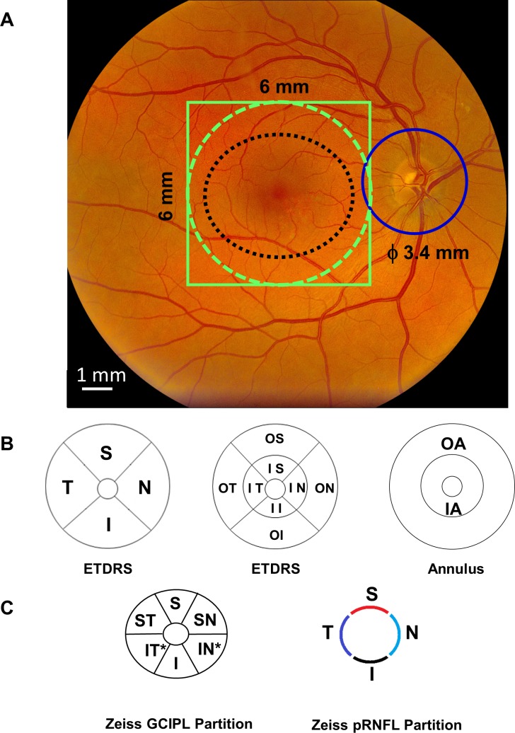
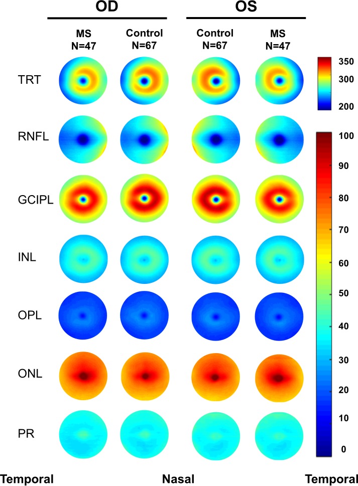
Ninety-four eyes of 47 relapsing-remitting MS patients without history of optic neuritis were imaged using optical coherence tomography and compared with 134 eyes of 67 healthy subjects. Volumetric data centered on the fovea were segmented to obtain the thickness maps of six intraretinal layers. The thickness measurements partitioned using the Early Treatment Diabetic Retinopathy Study (ETDRS) partition were correlated to the Expanded Disability State Scale (EDSS) and Sloan low contrast visual acuity (LCVA). The receiver-operating characteristics (ROC) curves were calculated to obtain the area under the ROC curves (AUCs).
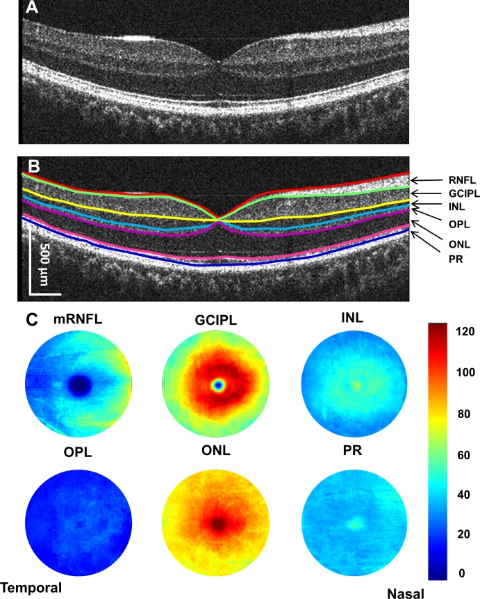
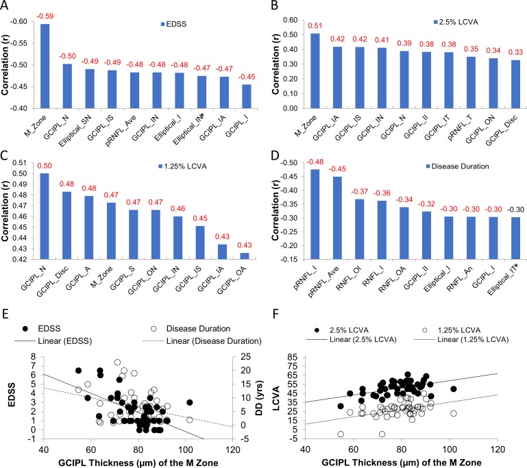
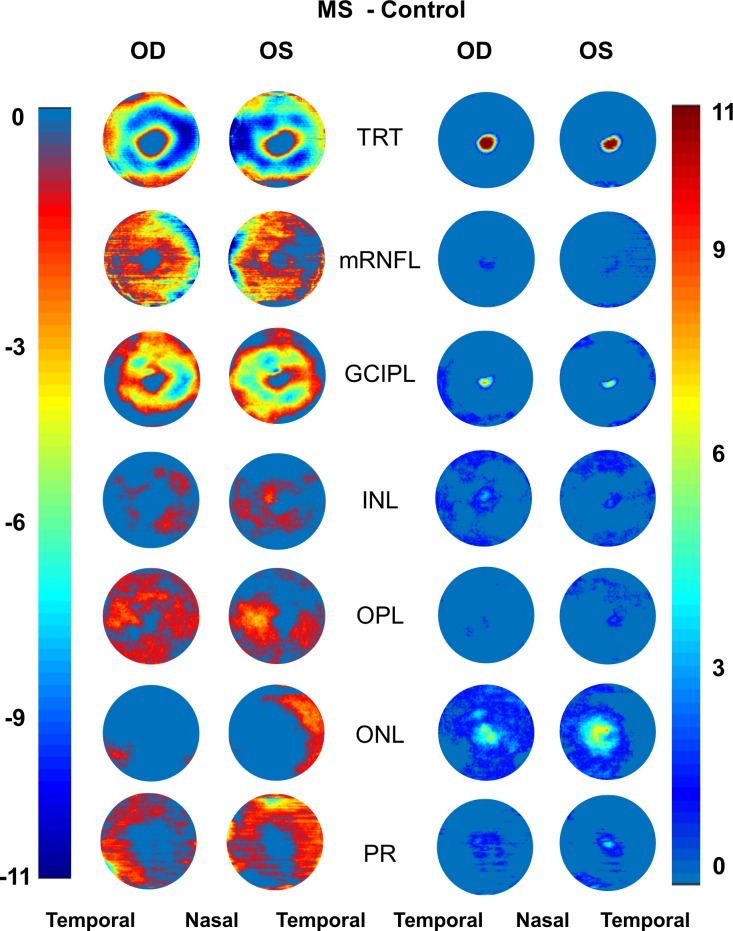
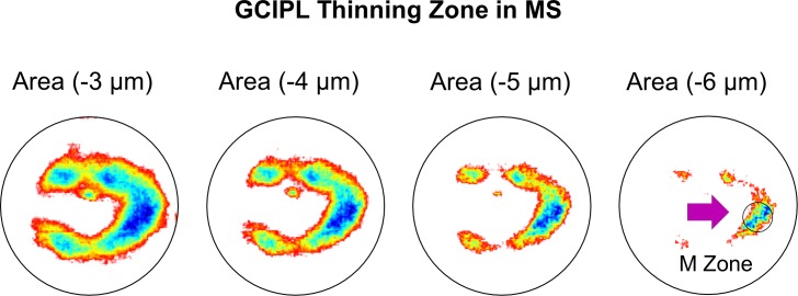
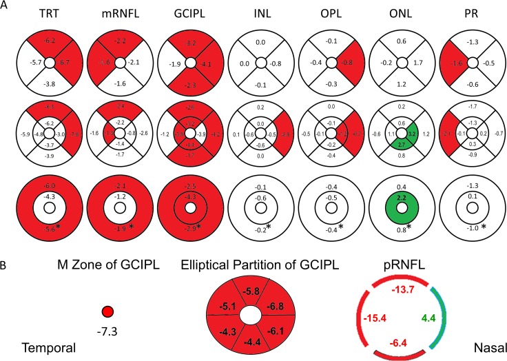
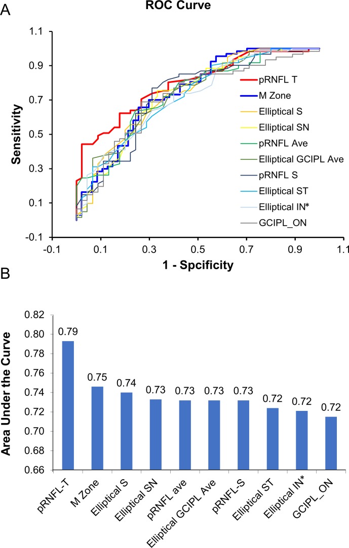
The ganglion cell-inner plexiform layer (GCIPL) showed horseshoe-like thickness reduction profoundly at the nasal sector. The most profound thickness reduction zone (circular area, diameter = 1 mm) was located at 2 mm in the nasal sector and 0.4 mm inferior from the fovea, named the "M zone." The thickness reduction of the M zone was -7.3 μm in MS eyes, which was the most profound alteration, compared to any ETDRS sectors. The AUC of the M zone was 0.75. The relationship between the thickness of the M zone and EDSS (r = -0.59, P < 0.001) or 2.5% LCVA (r = 0.51, P < 0.001) were ranked as the strongest relation compared to any ETDRS sectors.
This is the first study, to our knowledge, to visualize focal thickness alteration of GCIPL and reveal its relationship to visual function and disability in patients with MS without history of optic neuritis.
本研究旨在可视化多发性硬化症(MS)患者视网膜内各层的厚度分布模式及其与临床表现的相关性。
对 47 例无视神经炎病史的复发缓解型 MS 患者的 94 只眼进行光学相干断层扫描成像,并与 67 例健康对照者的 134 只眼进行比较。以黄斑为中心进行容积数据分割,获得 6 层视网膜内厚度图。采用早期治疗糖尿病视网膜病变研究(ETDRS)分区对厚度测量进行分区,并与扩展残疾状态量表(EDSS)和 Sloan 低对比视力(LCVA)相关联。计算受试者工作特征(ROC)曲线以获得 ROC 曲线下面积(AUC)。
神经节细胞-内丛状层(GCIPL)在鼻侧扇形变薄。最明显的厚度减小区域(圆形区域,直径= 1mm)位于鼻侧 2mm 处和黄斑下 0.4mm 处,命名为“M 区”。MS 眼 M 区的厚度减少为-7.3μm,与任何 ETDRS 区相比,这是最明显的改变。M 区的 AUC 为 0.75。M 区厚度与 EDSS(r=-0.59,P<0.001)或 2.5% LCVA(r=-0.51,P<0.001)之间的关系均强于任何 ETDRS 区。
据我们所知,这是第一项研究,旨在可视化无视神经炎病史的 MS 患者 GCIPL 的局灶性厚度改变,并揭示其与视觉功能和残疾的关系。