Wang Chao, Dang Yalong, Loewen Ralitsa T, Waxman Susannah, Shah Priyal, Xia Xiaobo, Loewen Nils A
Department of Ophthalmology, University of Pittsburgh Medical Center, Pittsburgh, PA, USA.
Department of Ophthalmology, Xiangya Hospital, Central South University, Changsha, Hunan, China.
Graefes Arch Clin Exp Ophthalmol. 2019 Jun;257(6):1217-1230. doi: 10.1007/s00417-019-04300-7. Epub 2019 Mar 28.
Dysfunction of the trabecular meshwork (TM) in pigmentary glaucoma contributes to increased aqueous humor outflow resistance and intraocular pressure. In this study, we investigated the effect of pigment dispersion on trabecular meshwork cells.
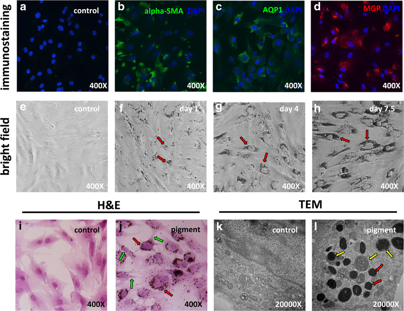
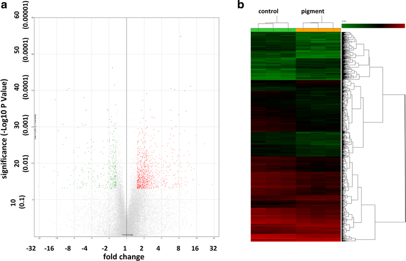
Porcine TM cells from ab interno trabeculectomy specimens were exposed to pigment dispersion, then, analyzed for changes in morphology, immunostaining, and ultrastructure. Their abilities to phagocytose migrate, and contraction was quantified. An expression microarray, using 23,937 probes, and a pathway analysis were performed.
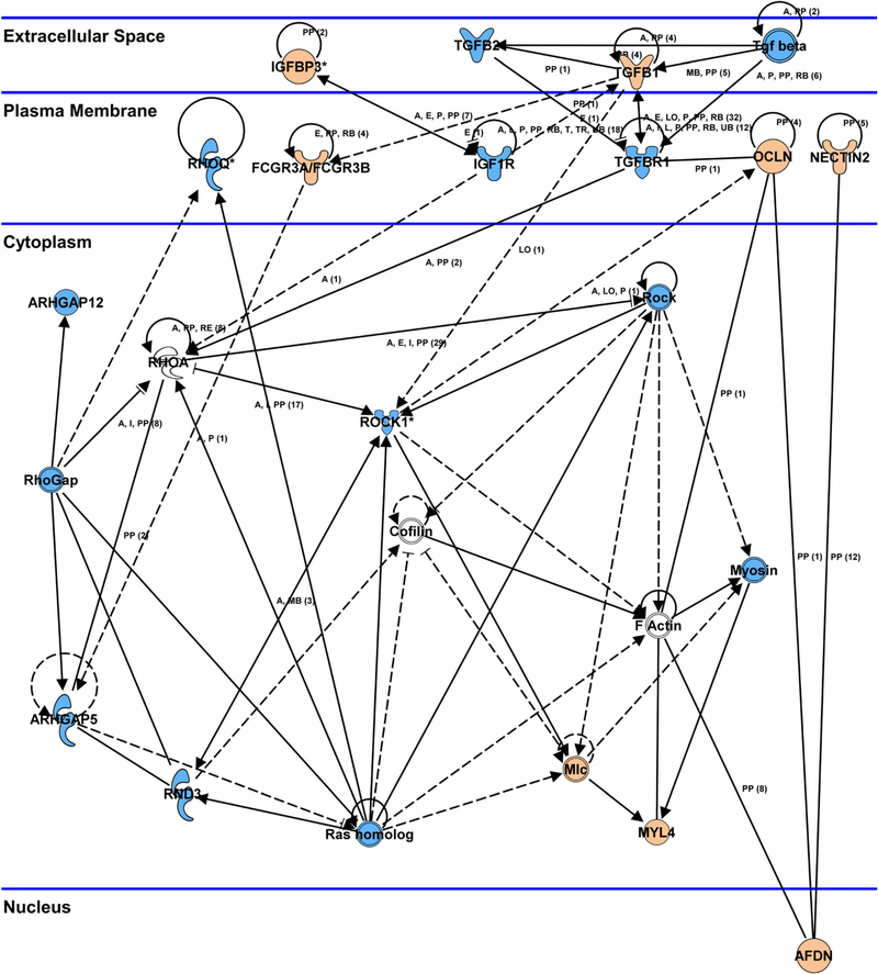
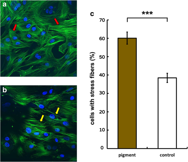
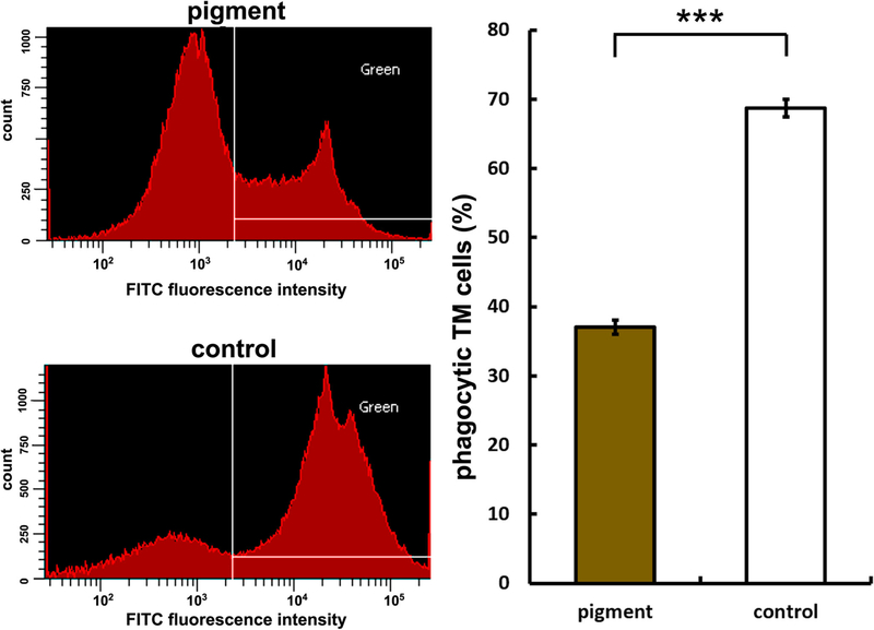
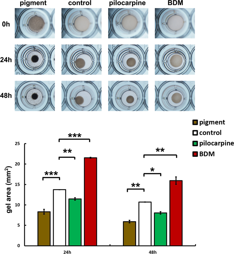
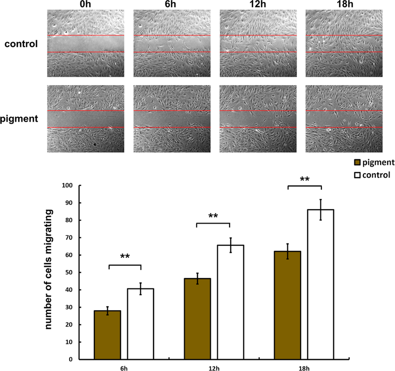
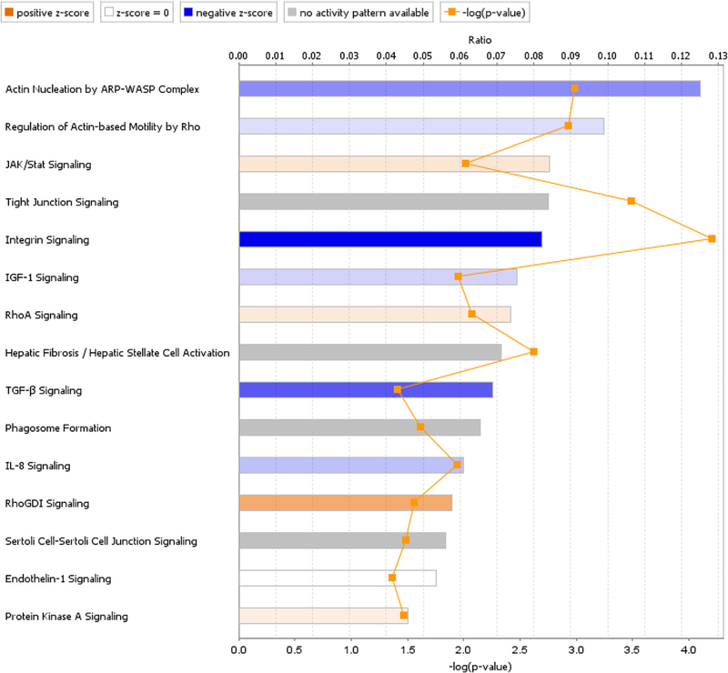
Stress fiber formation was increased in the pigment dispersion group (P) (60.1 ± 0.3%, n = 10) compared to control (C) (38.4 ± 2.5%, n = 11, p < 0.001). Phagocytosis declined (number of cells with microspheres in P = 37.0 ± 1.1% and in C = 68.7 ± 1.3%, n = 3, p < 0.001) and migration was reduced after 6 h (cells within the visual field over 6 h in P = 28.0.1 ± 2.3 (n = 12) and in C = 40.6 ± 3.3 (n = 13), p < 0.01). Pigment induced contraction at 24 h onwards (p < 0.01). Microarray analysis revealed that Rho signaling was central to these responses.
Exposure of TM cells to pigment dispersion resulted in reduced phagocytosis and migration, as well as increased stress fiber formation and cell contraction. The Rho signaling pathway played a central and early role, suggesting that its inhibitors could be used as a specific intervention in treatment of pigmentary glaucoma.
色素性青光眼患者小梁网(TM)功能障碍会导致房水流出阻力增加和眼压升高。在本研究中,我们调查了色素颗粒分散对小梁网细胞的影响。
将来自内路小梁切除术标本的猪TM细胞暴露于色素颗粒分散环境中,然后分析其形态、免疫染色和超微结构的变化。对其吞噬、迁移和收缩能力进行量化。使用23937个探针进行表达微阵列分析和通路分析。
与对照组(C)(38.4±2.5%,n = 11,p < 0.001)相比,色素颗粒分散组(P)(60.1±0.3%,n = 10)应力纤维形成增加。吞噬作用下降(P组含微球的细胞数为37.0±1.1%,C组为68.7±1.3%,n = 3,p < 0.001),6小时后迁移能力降低(P组6小时内视野内细胞数为28.0±2.3(n = 12),C组为40.6±3.3(n = 13),p < 0.01)。色素在24小时后开始诱导细胞收缩(p < 0.01)。微阵列分析显示Rho信号通路是这些反应的核心。
TM细胞暴露于色素颗粒分散环境中会导致吞噬和迁移能力降低,以及应力纤维形成和细胞收缩增加。Rho信号通路发挥了核心且早期的作用,这表明其抑制剂可作为色素性青光眼治疗的一种特异性干预手段。