Jansen Ilaria, Lucas Marit, Savci-Heijink C Dilara, Meijer Sybren L, Liem Esmee I M L, de Boer Onno J, van Leeuwen Ton G, Marquering Henk A, de Bruin Daniel M
Department of Biomedical Engineering and Physics, Amsterdam UMC, University of Amsterdam, Amsterdam, The Netherlands.
Department of Urology, Amsterdam UMC, University of Amsterdam, Amsterdam, The Netherlands.
Diagn Pathol. 2019 Mar 28;14(1):25. doi: 10.1186/s13000-019-0803-7.
Histopathological analysis is the cornerstone in bladder cancer (BCa) diagnosis. These analysis suffer from a moderate observer agreement in the staging of bladder cancer. Three-dimensional reconstructions have the potential to support the pathologists in visualizing spatial arrangements of structures, which may improve the interpretation of specimen. The aim of this study is to present three-dimensional (3D) reconstructions of histology images.
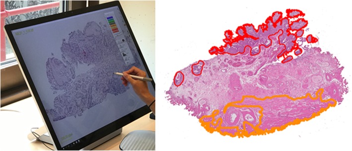
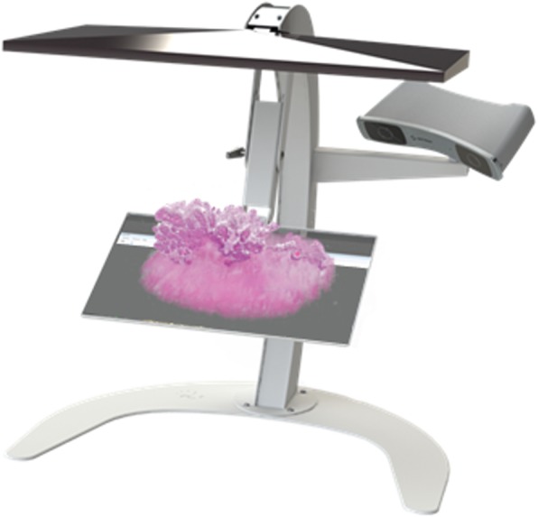
En-bloc specimens of transurethral bladder tumour resections were formalin fixed and paraffin embedded. Specimens were cut into sections of 4 μm and stained with Hematoxylin and Eosin (H&E). With a Phillips IntelliSite UltraFast scanner, glass slides were digitized at 20x magnification. The digital images were aligned by performing rigid and affine image alignment. The tumour and the muscularis propria (MP) were manually delineated to create 3D segmentations. In conjunction with a 3D display, the results were visualized with the Vesalius3D interactive visualization application for a 3D workstation.
En-bloc resection was performed in 21 BCa patients. Per case, 26-30 sections were included for the reconstruction into a 3D volume. Five cases were excluded due to export problems, size of the dataset or condition of the tissue block. Qualitative evaluation suggested an accurate registration for 13 out of 16 cases. The segmentations allowed full 3D visualization and evaluation of the spatial relationship of the BCa tumour and the MP.
Digital scanning of en-bloc resected specimens allows a full-fledged 3D reconstruction and analysis and has a potential role to support pathologists in the staging of BCa.
组织病理学分析是膀胱癌(BCa)诊断的基石。然而,这些分析在膀胱癌分期方面的观察者间一致性一般。三维重建有潜力辅助病理学家可视化结构的空间排列,这可能会改善对标本的解读。本研究的目的是展示组织学图像的三维(3D)重建。
经尿道膀胱肿瘤切除术的整块标本用福尔马林固定并石蜡包埋。标本切成4μm厚的切片,并用苏木精和伊红(H&E)染色。使用飞利浦IntelliSite UltraFast扫描仪,以20倍放大倍数对载玻片进行数字化处理。通过执行刚性和仿射图像配准来对齐数字图像。手动勾勒肿瘤和固有肌层(MP)以创建三维分割。结合三维显示,使用Vesalius3D交互式可视化应用程序在三维工作站上对结果进行可视化。
对21例BCa患者进行了整块切除。每例纳入26 - 30个切片用于重建三维体积。由于导出问题、数据集大小或组织块状况,排除了5例。定性评估表明16例中有13例配准准确。这些分割允许对BCa肿瘤和MP的空间关系进行完整的三维可视化和评估。
整块切除标本的数字扫描允许进行全面的三维重建和分析,并且在支持病理学家进行BCa分期方面具有潜在作用。