• 文献检索
  • 文档翻译
  • 深度研究
  • 学术资讯
  • Suppr Zotero 插件Zotero 插件
  • 邀请有礼
  • 套餐&价格
  • 历史记录
应用&插件
Suppr Zotero 插件Zotero 插件浏览器插件Mac 客户端Windows 客户端微信小程序
定价
高级版会员购买积分包购买API积分包
服务
文献检索文档翻译深度研究API 文档MCP 服务
关于我们
关于 Suppr公司介绍联系我们用户协议隐私条款
关注我们

Suppr 超能文献

核心技术专利:CN118964589B侵权必究
粤ICP备2023148730 号-1Suppr @ 2026

文献检索

告别复杂PubMed语法,用中文像聊天一样搜索,搜遍4000万医学文献。AI智能推荐,让科研检索更轻松。

立即免费搜索

文件翻译

保留排版,准确专业,支持PDF/Word/PPT等文件格式,支持 12+语言互译。

免费翻译文档

深度研究

AI帮你快速写综述,25分钟生成高质量综述,智能提取关键信息,辅助科研写作。

立即免费体验

开发一种用于非小细胞肺癌 PET-CT 图像与全组织切片病理的三维相关性分析的方法。

Developing a methodology for three-dimensional correlation of PET-CT images and whole-mount histopathology in non-small-cell lung cancer.

机构信息

Radiation Medicine Program, Princess Margaret Hospital, University Health Network, Toronto, ON.

出版信息

Curr Oncol. 2008 Oct;15(5):62-9. doi: 10.3747/co.v15i5.349.

DOI:10.3747/co.v15i5.349
PMID:19008992
原文链接:https://pmc.ncbi.nlm.nih.gov/articles/PMC2582510/
Abstract

BACKGROUND

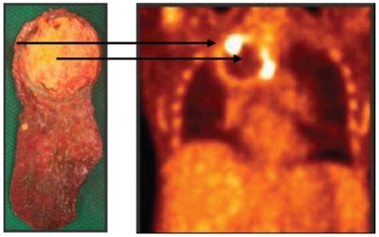
Understanding the three-dimensional (3D) volumetric relationship between imaging and functional or histopathologic heterogeneity of tumours is a key concept in the development of image-guided radiotherapy. Our aim was to develop a methodologic framework to enable the reconstruction of resected lung specimens containing non-small-cell lung cancer (NSCLC), to register the result in 3D with diagnostic imaging, and to import the reconstruction into a radiation treatment planning system.

METHODS AND RESULTS

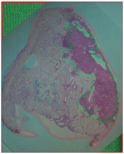
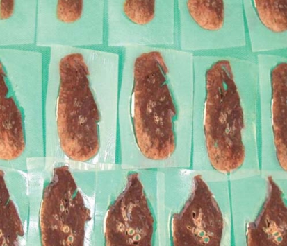
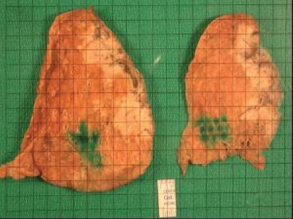
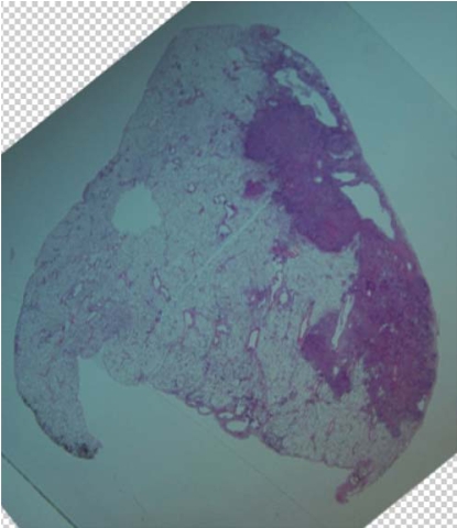
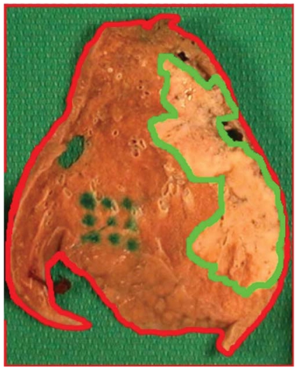
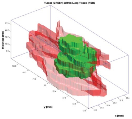
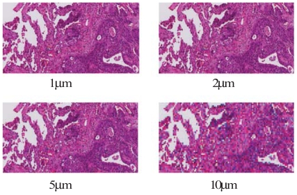
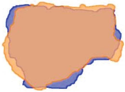
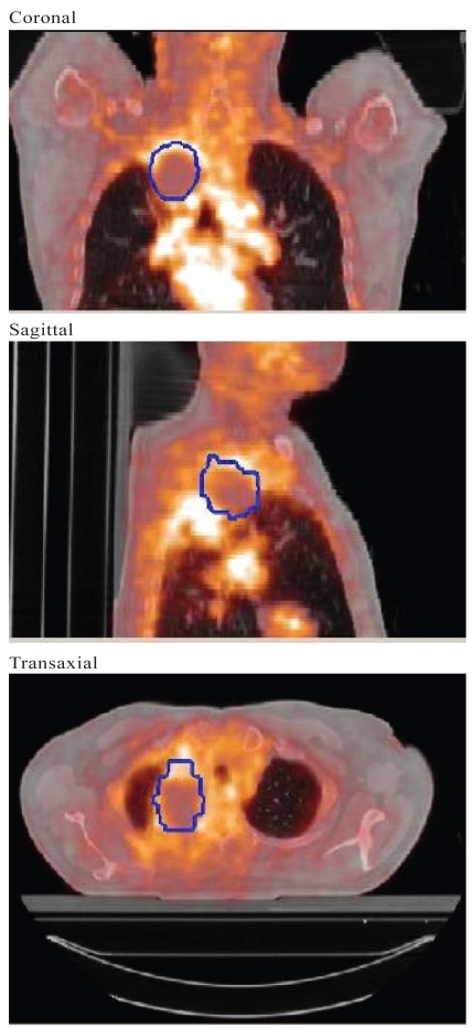
We recruited 12 patients for an investigation of radiology-pathology correlation (RPC) in nsclc. Before resection, imaging by positron emission tomography (PET) or computed tomography (CT) was obtained. Resected specimens were formalin-fixed for 1-24 hours before sectioning at 3-mm to 10-mm intervals. To try to retain the original shape, we embedded the specimens in agar before sectioning. Consecutive sections were laid out for photography and manually adjusted to maintain shape. Following embedding, the tissue blocks underwent whole-mount sectioning (4-mum sections) and staining with hematoxylin and eosin. Large histopathology slides were used to whole-mount entire sections for digitization. The correct sequence was maintained to assist in subsequent reconstruction. Using Photoshop (Adobe Systems Incorporated, San Jose, CA, U.S.A.), contours were placed on the photographic images to represent the external borders of the section and the extent of macroscopic disease. Sections were stacked in sequence and manually oriented in Photoshop. The macroscopic tumour contours were then transferred to MATLAB (The Mathworks, Natick, MA, U.S.A.) and stacked, producing 3D surface renderings of the resected specimen and embedded gross tumour. To evaluate the microscopic extent of disease, customized "tile-based" and commercial confocal panoramic laser scanning (TISSUEscope: Biomedical Photometrics, Waterloo, ON) systems were used to generate digital images of whole-mount histopathology sections. Using the digital whole-mount images and imaging software, we contoured the gross and microscopic extent of disease. Two methods of registering pathology and imaging were used. First, selected pet and ct images were transferred into Photoshop, where they were contoured, stacked, and reconstructed. After importing the pathology and the imaging contours to MATLAB, the contours were reconstructed, manually rotated, and rigidly registered. In the second method, MATLAB tumour renderings were exported to a software platform for manual registration with the original pet and ct images in multiple planes. Data from this software platform were then exported to the Pinnacle radiation treatment planning system in DICOM (Digital Imaging and Communications in Medicine) format.

CONCLUSIONS

There is no one definitive method for 3D volumetric RPC in nsclc. An innovative approach to the 3D reconstruction of resected nsclc specimens incorporates agar embedding of the specimen and whole-mount digital histopathology. The reconstructions can be rigidly and manually registered to imaging modalities such as ct and pet and exported to a radiation treatment planning system.

摘要

背景

了解成像与肿瘤功能或组织病理学异质性的三维(3D)体积关系是图像引导放疗发展的关键概念。我们的目的是开发一种方法学框架,使包含非小细胞肺癌(NSCLC)的切除肺标本能够重建,并在 3D 中与诊断成像进行配准,并将重建导入放射治疗计划系统。

方法和结果

我们招募了 12 名患者进行 NSCLC 的放射病理学相关性(RPC)研究。在切除前,通过正电子发射断层扫描(PET)或计算机断层扫描(CT)进行成像。切除的标本在福尔马林固定 1-24 小时后,以 3-10mm 的间隔进行切片。为了尽量保持原形,我们在切片前将标本嵌入琼脂中。连续切片用于摄影,并手动调整以保持形状。包埋后,组织块进行全切片(4μm 切片)和苏木精-伊红染色。使用大的组织病理学幻灯片对整个切片进行全载片数字化。保持正确的顺序有助于随后的重建。使用 Photoshop(Adobe Systems Incorporated,美国圣何塞),在摄影图像上放置轮廓以代表切片的外部边界和大体疾病的范围。切片按顺序堆叠并在 Photoshop 中手动定向。然后将大体肿瘤轮廓转移到 MATLAB(Mathworks,美国马萨诸塞州纳蒂克)并堆叠,生成切除标本和嵌入大体肿瘤的 3D 表面渲染。为了评估疾病的微观范围,使用定制的“基于瓦片”和商业共焦全景激光扫描(TISSUEscope:Biomedical Photometrics,安大略省滑铁卢)系统生成全载片组织病理学切片的数字图像。使用数字全载片图像和成像软件,我们对大体和微观疾病范围进行了轮廓描绘。使用了两种配准病理学和成像的方法。首先,将选定的 PET 和 CT 图像传输到 Photoshop 中,在其中进行轮廓绘制、堆叠和重建。将病理学和成像轮廓导入 MATLAB 后,重建轮廓,手动旋转并刚性配准。在第二种方法中,将 MATLAB 肿瘤渲染导出到一个软件平台,用于在多个平面上与原始的 PET 和 CT 图像进行手动配准。然后,从该软件平台导出数据到 Pinnacle 放射治疗计划系统的 DICOM(医学数字成像和通信)格式。

结论

在 NSCLC 中,没有一种确定的 3D 容积 RPC 方法。一种对切除的 NSCLC 标本进行 3D 重建的创新方法是将标本嵌入琼脂和全载片数字化组织病理学。重建可以与 CT 和 PET 等成像方式刚性和手动配准,并导出到放射治疗计划系统。

https://cdn.ncbi.nlm.nih.gov/pmc/blobs/1706/2582510/adcc5d4fef2b/co15-5-225e62f10.jpg
https://cdn.ncbi.nlm.nih.gov/pmc/blobs/1706/2582510/34d2459eb166/co15-5-225e62f1.jpg
https://cdn.ncbi.nlm.nih.gov/pmc/blobs/1706/2582510/c111f07b08e5/co15-5-225e62f2.jpg
https://cdn.ncbi.nlm.nih.gov/pmc/blobs/1706/2582510/067019691753/co15-5-225e62f3.jpg
https://cdn.ncbi.nlm.nih.gov/pmc/blobs/1706/2582510/1ca09c28c453/co15-5-225e62f4.jpg
https://cdn.ncbi.nlm.nih.gov/pmc/blobs/1706/2582510/5c5f6b998240/co15-5-225e62f5.jpg
https://cdn.ncbi.nlm.nih.gov/pmc/blobs/1706/2582510/ca5c5cf7313b/co15-5-225e62f6.jpg
https://cdn.ncbi.nlm.nih.gov/pmc/blobs/1706/2582510/148f88f501d1/co15-5-225e62f7.jpg
https://cdn.ncbi.nlm.nih.gov/pmc/blobs/1706/2582510/e43dc81d57fa/co15-5-225e62f8.jpg
https://cdn.ncbi.nlm.nih.gov/pmc/blobs/1706/2582510/5949b8016c4a/co15-5-225e62f9.jpg
https://cdn.ncbi.nlm.nih.gov/pmc/blobs/1706/2582510/adcc5d4fef2b/co15-5-225e62f10.jpg

相似文献

1
Developing a methodology for three-dimensional correlation of PET-CT images and whole-mount histopathology in non-small-cell lung cancer.开发一种用于非小细胞肺癌 PET-CT 图像与全组织切片病理的三维相关性分析的方法。
Curr Oncol. 2008 Oct;15(5):62-9. doi: 10.3747/co.v15i5.349.
2
Improving superficial target delineation in radiation therapy with endoscopic tracking and registration.利用内镜跟踪和配准技术改善放射治疗中的表面靶区勾画。
Med Phys. 2011 Dec;38(12):6458-68. doi: 10.1118/1.3658569.
3
Feasibility of [18F]FDG-PET and coregistered CT on clinical target volume definition of advanced non-small cell lung cancer.[18F]氟代脱氧葡萄糖正电子发射断层扫描([18F]FDG-PET)及配准CT在晚期非小细胞肺癌临床靶区定义中的可行性
Q J Nucl Med Mol Imaging. 2005 Sep;49(3):259-66.
4
A method for accurate spatial registration of PET images and histopathology slices.一种用于PET图像与组织病理学切片精确空间配准的方法。
EJNMMI Res. 2015 Dec;5(1):64. doi: 10.1186/s13550-015-0138-7. Epub 2015 Nov 14.
5
Display of positron emission tomography with Cadplan.使用Cadplan的正电子发射断层扫描显示。
Australas Phys Eng Sci Med. 2002 Jul;25(2):67-77. doi: 10.1007/BF03178468.
6
Comparison of positron emission tomography (PET) and computed tomography (CT) for better target volume definition in radiation therapy planning.正电子发射断层扫描(PET)和计算机断层扫描(CT)在放疗计划中更好地定义靶区体积的比较。
Clin Transl Oncol. 2010 May;12(5):367-73. doi: 10.1007/s12094-010-0518-4.
7
Automated detection, delineation and quantification of whole-body bone metastasis using FDG-PET/CT images.使用 FDG-PET/CT 图像自动检测、勾画和定量全身骨转移。
Phys Eng Sci Med. 2023 Jun;46(2):851-863. doi: 10.1007/s13246-023-01258-z. Epub 2023 May 1.
8
Validation of imaging with pathology in laryngeal cancer: accuracy of the registration methodology.喉癌影像学与病理学验证:配准方法的准确性。
Int J Radiat Oncol Biol Phys. 2012 Feb 1;82(2):e289-98. doi: 10.1016/j.ijrobp.2011.05.004. Epub 2011 Jun 28.
9
Impact of FDG-PET on radiation therapy volume delineation in non-small-cell lung cancer.18F-氟脱氧葡萄糖正电子发射断层扫描对非小细胞肺癌放射治疗靶区勾画的影响
Int J Radiat Oncol Biol Phys. 2004 May 1;59(1):78-86. doi: 10.1016/j.ijrobp.2003.10.044.
10
Comparison of imaging-based gross tumor volume and pathological volume determined by whole-mount serial sections in primary cervical cancer.基于影像学的大体肿瘤体积与全子宫连续切片病理测量原发性宫颈癌肿瘤体积的比较。
Onco Targets Ther. 2013 Jul 19;6:917-23. doi: 10.2147/OTT.S43264. Print 2013.

引用本文的文献

1
Prospective validation of craniocaudal tumour size on MR imaging compared to histoPAthology in patients with uterine cervical cancer: The MPAC study.子宫颈癌患者中,与组织病理学相比,磁共振成像上肿瘤头尾径大小的前瞻性验证:MPAC研究。
Clin Transl Radiat Oncol. 2019 Jun 15;18:9-15. doi: 10.1016/j.ctro.2019.06.004. eCollection 2019 Sep.
2
Analysis of Risk Factors for Lymphatic Metastasis in Endometrial Carcinoma and Utility of Three-Dimensional Magnetic Resonance Imaging in Gynecology.子宫内膜癌淋巴转移的危险因素分析及三维磁共振成像在妇科中的应用
World J Oncol. 2018 Jun;9(3):74-79. doi: 10.14740/wjon1106w. Epub 2018 Jun 26.
3

本文引用的文献

1
Correlation of intra-tumour heterogeneity on 18F-FDG PET with pathologic features in non-small cell lung cancer: a feasibility study.18F-FDG PET上肿瘤内异质性与非小细胞肺癌病理特征的相关性:一项可行性研究
Radiother Oncol. 2008 Apr;87(1):55-8. doi: 10.1016/j.radonc.2008.02.002. Epub 2008 Mar 6.
2
Feasibility of pathology-correlated lung imaging for accurate target definition of lung tumors.用于准确界定肺肿瘤靶区的病理学相关肺部成像的可行性
Int J Radiat Oncol Biol Phys. 2007 Sep 1;69(1):267-75. doi: 10.1016/j.ijrobp.2007.04.065.
3
Clinicopathologic analysis of microscopic extension in lung adenocarcinoma: defining clinical target volume for radiotherapy.
Will the mininvasive approach challenge the old paradigms in oral cancer surgery?
微创方法会挑战口腔癌手术的旧有模式吗?
Eur Arch Otorhinolaryngol. 2017 Mar;274(3):1279-1289. doi: 10.1007/s00405-016-4221-0. Epub 2016 Aug 4.
4
Detection of aberrant hippocampal mossy fiber connections: Ex vivo mesoscale diffusion MRI and microtractography with histological validation in a patient with uncontrolled temporal lobe epilepsy.异常海马苔藓纤维连接的检测:一名颞叶癫痫未控制患者的离体中尺度扩散磁共振成像和微观纤维束成像及组织学验证
Hum Brain Mapp. 2016 Feb;37(2):780-95. doi: 10.1002/hbm.23066. Epub 2015 Nov 27.
5
Comparison of imaging-based gross tumor volume and pathological volume determined by whole-mount serial sections in primary cervical cancer.基于影像学的大体肿瘤体积与全子宫连续切片病理测量原发性宫颈癌肿瘤体积的比较。
Onco Targets Ther. 2013 Jul 19;6:917-23. doi: 10.2147/OTT.S43264. Print 2013.
6
Automatic Segmentation of Lung Carcinoma Using 3D Texture Features in 18-FDG PET/CT.利用18-FDG PET/CT中的3D纹理特征自动分割肺癌
Int J Mol Imaging. 2013;2013:980769. doi: 10.1155/2013/980769. Epub 2013 Feb 26.
7
The value of large sections in surgical pathology.大体标本在外科病理学中的价值。
Int J Breast Cancer. 2012;2012:785947. doi: 10.1155/2012/785947. Epub 2012 Nov 21.
8
New strategy for automatic tumor segmentation by adaptive thresholding on PET/CT images.基于 PET/CT 图像自适应阈值的肿瘤自动分割新策略。
J Appl Clin Med Phys. 2012 Sep 6;13(5):3875. doi: 10.1120/jacmp.v13i5.3875.
9
Determination of infarct size in ex vivo swine hearts by multidetector computed tomography using gadolinium as contrast medium.应用钆造影剂的多排 CT 对离体猪心梗死面积的测定。
Invest Radiol. 2012 May;47(5):277-83. doi: 10.1097/RLI.0b013e31823fabc6.
10
Comparative methods for PET image segmentation in pharyngolaryngeal squamous cell carcinoma.头颈部鳞癌正电子发射断层扫描图像分割的比较方法。
Eur J Nucl Med Mol Imaging. 2012 May;39(5):881-91. doi: 10.1007/s00259-011-2053-0.
肺腺癌微小浸润的临床病理分析:确定放射治疗的临床靶区
Int J Radiat Oncol Biol Phys. 2007 Oct 1;69(2):334-41. doi: 10.1016/j.ijrobp.2007.03.023. Epub 2007 Jun 14.
4
Whole-specimen histopathology: a method to produce whole-mount breast serial sections for 3-D digital histopathology imaging.全标本组织病理学:一种制作用于三维数字组织病理学成像的乳腺连续整装切片的方法。
Histopathology. 2007 Jan;50(2):232-42. doi: 10.1111/j.1365-2559.2006.02561.x.
5
Design and characterization of a digital image acquisition system for whole-specimen breast histopathology.用于全标本乳腺组织病理学的数字图像采集系统的设计与特性分析
Phys Med Biol. 2006 Oct 21;51(20):5089-103. doi: 10.1088/0031-9155/51/20/002. Epub 2006 Sep 22.
6
Non-rigid image registration: theory and practice.非刚性图像配准:理论与实践
Br J Radiol. 2004;77 Spec No 2:S140-53. doi: 10.1259/bjr/25329214.
7
Tumor volume in pharyngolaryngeal squamous cell carcinoma: comparison at CT, MR imaging, and FDG PET and validation with surgical specimen.咽喉部鳞状细胞癌的肿瘤体积:CT、磁共振成像及氟代脱氧葡萄糖正电子发射断层显像的比较及手术标本验证
Radiology. 2004 Oct;233(1):93-100. doi: 10.1148/radiol.2331030660. Epub 2004 Aug 18.
8
[A comparative study on radiology and pathology target volume in non-small-cell lung cancer].非小细胞肺癌放射学与病理学靶区的对比研究
Zhonghua Zhong Liu Za Zhi. 2003 Nov;25(6):566-8.
9
Wedge resection margin distances and residual adenocarcinoma in lobectomy specimens.肺叶切除标本中的楔形切除切缘距离和残余腺癌
Am J Clin Pathol. 2003 Nov;120(5):720-4. doi: 10.1309/P47F-YW5U-4CRQ-0WFE.
10
Definition of gross tumor volume in lung cancer: inter-observer variability.肺癌中肿瘤大体体积的定义:观察者间的变异性。
Radiother Oncol. 2002 Jan;62(1):37-49. doi: 10.1016/s0167-8140(01)00453-4.