Casey Eye Institute, Oregon Health & Science University, Portland, Oregon, United States.
Department of Physiology & Pharmacology, Oregon Health & Science University, Portland, Oregon, United States.
Invest Ophthalmol Vis Sci. 2019 Mar 1;60(4):1275-1285. doi: 10.1167/iovs.18-24398.
We determine if monomethyl fumarate (MMF) can protect the retina in mice subjected to light-induced retinopathy (LIR).
Albino BALB/c mice were intraperitoneally injected with 50 to 100 mg/kg MMF before or after exposure to bright white light (10,000 lux) for 1 hour. Seven days after light exposure, retinal structure and function were evaluated by optical coherence tomography (OCT) and electroretinography (ERG), respectively. Retinal histology also was performed to evaluate photoreceptor loss. Expression levels of Hcar2 and markers of microglia activation were measured by quantitative PCR (qPCR) in the neural retina with and without microglia depletion. At 24 hours after light exposure, retinal sections and whole mount retinas were stained with Iba1 to evaluate microglia status. The effect of MMF on the nuclear factor kB subunit 1 (NF-kB) and Nrf2 pathways was measured by qPCR and Western blot.
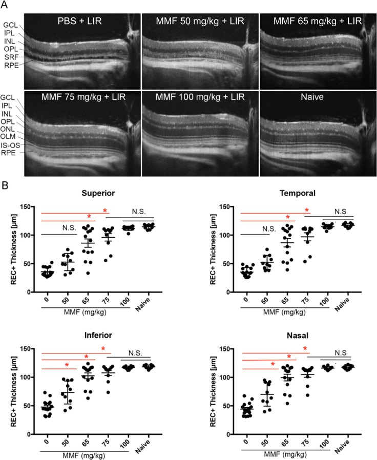
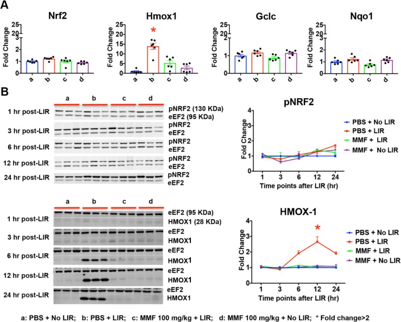
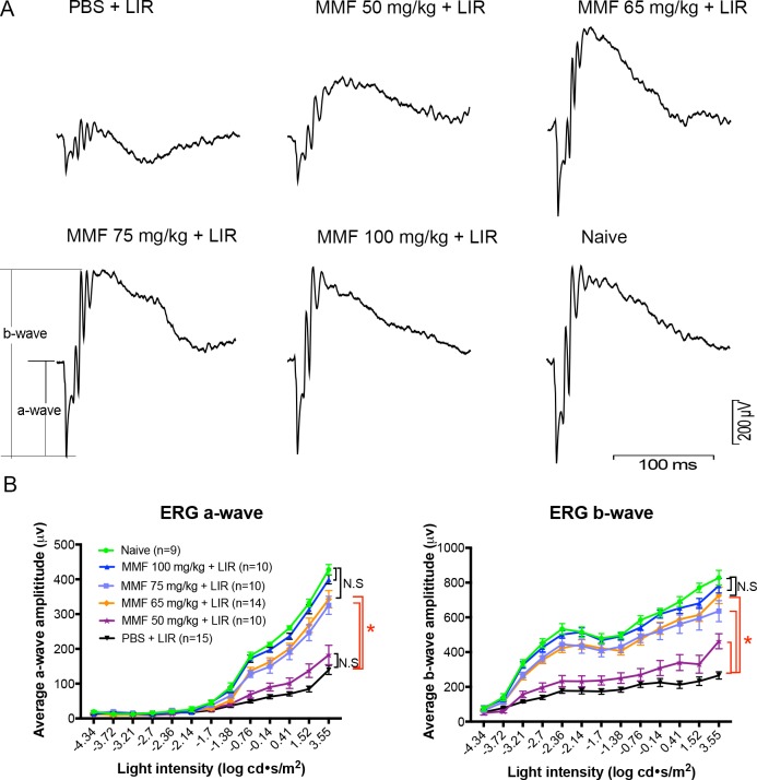
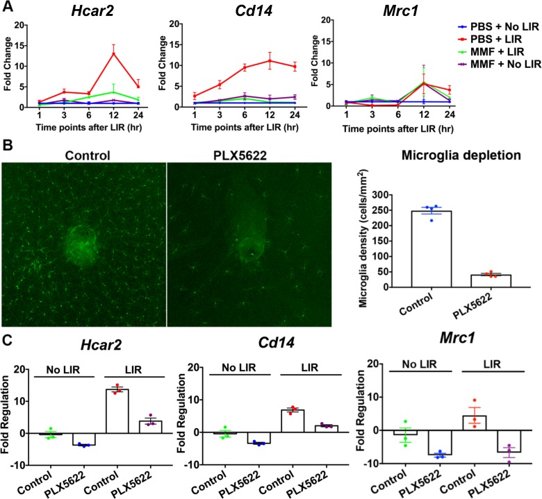
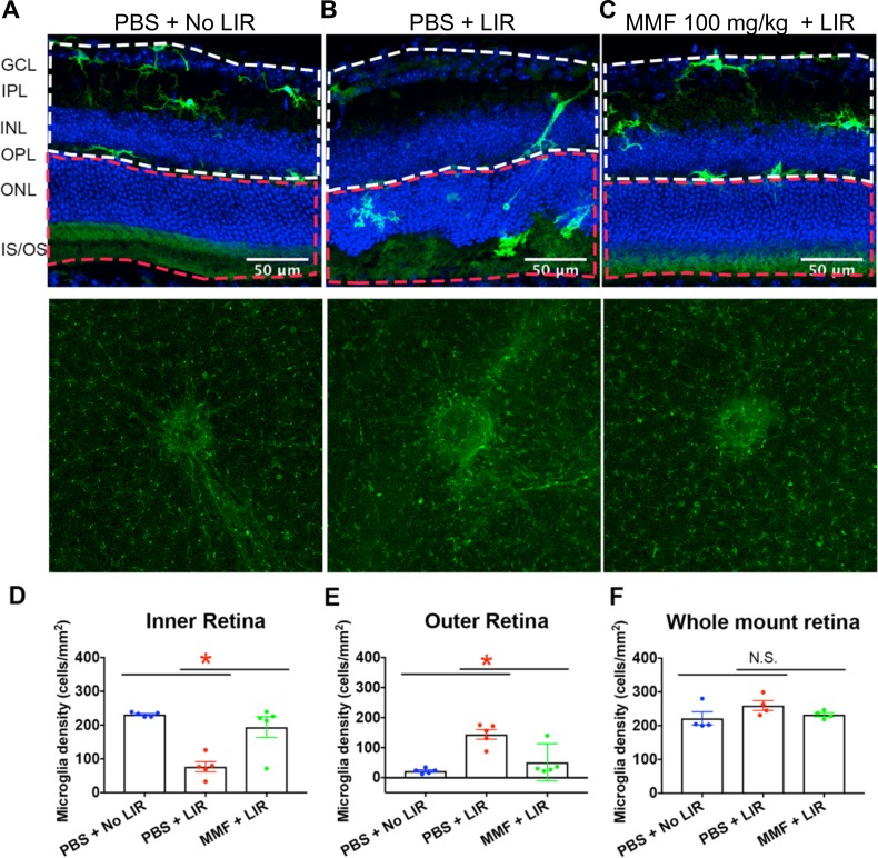
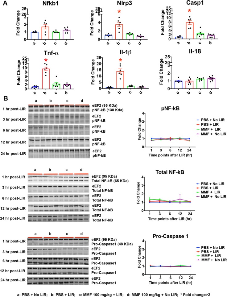
MMF administered before light exposure mediated dose-dependent neuroprotection in a mouse model of LIR. A single dose of 100 mg/kg MMF fully protected retinal structure and function without side effects. Expression of the Hcar2 receptor and the microglia marker Cd14 were upregulated by LIR, but suppressed by MMF. Depleting microglia reduced Hcar2 expression and its upregulation by LIR. Microglial activation, upregulation of proinflammatory genes (Nlrp3, Caspase1, Il-1β, Tnf-α), and upregulation of antioxidative stress genes (Hmox1) associated with LIR were mitigated by MMF treatment.
MMF can completely protect the retina from LIR in BALB/c mice. Expression of Hcar2, the receptor of MMF, is microglia-dependent in the neural retina. MMF-mediated neuroprotection was associated with attenuation of microglia activation, inflammation and oxidative stress in the retina.
我们旨在确定富马酸单甲酯(MMF)是否能保护光诱导的视网膜病变(LIR)小鼠的视网膜。
白化 BALB/c 小鼠在暴露于 10000 勒克斯明亮白光前或后腹腔内注射 50-100mg/kg MMF,暴露后 1 小时。光暴露 7 天后,分别通过光学相干断层扫描(OCT)和视网膜电图(ERG)评估视网膜结构和功能。视网膜组织学也用于评估光感受器损失。用定量 PCR(qPCR)在有和无小胶质细胞耗竭的神经视网膜中测量 Hcar2 的表达水平和小胶质细胞激活的标志物。在光暴露后 24 小时,用 Iba1 对视网膜切片和全视网膜铺片进行染色,以评估小胶质细胞状态。用 qPCR 和 Western blot 测定 MMF 对核因子 kB 亚单位 1(NF-kB)和 Nrf2 途径的影响。
在 LIR 小鼠模型中,MMF 预先给药介导剂量依赖性神经保护作用。单次 100mg/kg MMF 完全保护视网膜结构和功能,无副作用。Hcar2 受体和小胶质细胞标志物 Cd14 的表达在 LIR 后上调,但被 MMF 抑制。小胶质细胞耗竭降低了 LIR 引起的 Hcar2 表达及其上调。MMF 处理减轻了与 LIR 相关的小胶质细胞激活、促炎基因(Nlrp3、Caspase1、IL-1β、TNF-α)上调和抗氧化应激基因(Hmox1)上调。
MMF 可完全保护 BALB/c 小鼠的视网膜免受 LIR 的损害。MMF 的受体 Hcar2 的表达在神经视网膜中依赖于小胶质细胞。MMF 介导的神经保护作用与小胶质细胞激活、炎症和氧化应激的减轻有关。