Lorio Sara, Sambataro Fabio, Bertolino Alessandro, Draganski Bogdan, Dukart Juergen
Developmental Neurosciences, UCL Great Ormond Street Institute of Child Health, University College London, London, United Kingdom.
Roche Pharma and Early Development, Neuroscience, Ophthalmology and Rare Diseases, F. Hoffmann-La Roche Ltd., Basel, Switzerland.
Front Aging Neurosci. 2019 Mar 15;11:57. doi: 10.3389/fnagi.2019.00057. eCollection 2019.
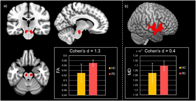
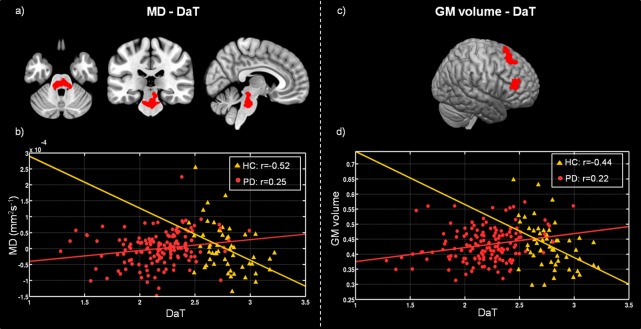
There is an increasing interest in identifying non-invasive biomarkers of disease severity and prognosis in idiopathic Parkinson's disease (PD). Dopamine-transporter SPECT (DAT-SPECT), diffusion tensor imaging (DTI), and structural magnetic resonance imaging (sMRI) provide unique information about the brain's neurotransmitter and microstructural properties. In this study, we evaluate the relative and combined capability of these imaging modalities to predict symptom severity and clinical progression in PD patients. To this end, we used MRI, SPECT, and clinical data of drug-naïve PD patients ( = 205, mean age 61 ± 10) and age-, sex-matched healthy controls ( = 105, mean age 58 ± 12) acquired at baseline. Moreover, we employed clinical data acquired at 1 year follow-up for PD patients with or without L-Dopa treatment in order to predict the progression symptoms severity. Voxel-based group comparisons and covariance analyses were applied to characterize baseline disease-related alterations for DAT-SPECT, DTI, and sMRI. Cortical and subcortical alterations in PD patients were found in all evaluated imaging modalities, in line with previously reported midbrain-striato-cortical network alterations. The combination of these imaging alterations was reliably linked to clinical severity and disease progression at 1 year follow-up in this patient population, providing evidence for the potential use of these modalities as imaging biomarkers for disease severity and prognosis that can be integrated into clinical trials.
人们对识别特发性帕金森病(PD)疾病严重程度和预后的非侵入性生物标志物的兴趣与日俱增。多巴胺转运体单光子发射计算机断层扫描(DAT-SPECT)、扩散张量成像(DTI)和结构磁共振成像(sMRI)提供了有关大脑神经递质和微观结构特性的独特信息。在本研究中,我们评估了这些成像方式预测PD患者症状严重程度和临床进展的相对及联合能力。为此,我们使用了初治PD患者(n = 205,平均年龄61±10岁)以及年龄和性别匹配的健康对照者(n = 105,平均年龄58±12岁)在基线时获取的MRI、SPECT和临床数据。此外,我们采用了在1年随访时获取的PD患者(无论是否接受左旋多巴治疗)的临床数据,以预测症状严重程度的进展情况。基于体素的组间比较和协方差分析用于表征DAT-SPECT、DTI和sMRI与疾病相关的基线改变。在所有评估的成像方式中均发现了PD患者的皮质和皮质下改变,这与先前报道的中脑-纹状体-皮质网络改变一致。在该患者群体中,这些成像改变的组合与1年随访时的临床严重程度和疾病进展可靠相关,为这些成像方式作为疾病严重程度和预后的成像生物标志物的潜在应用提供了证据,这些生物标志物可纳入临床试验。