Zhang Yu, Wu I-Wei, Tosun Duygu, Foster Eric, Schuff Norbert
Department of Veteran Affairs Medical Center, San Francisco, California, United States of America.
Department of Radiology and Biomedical Imaging, University of California, San Francisco, California, United States of America.
PLoS One. 2016 Oct 31;11(10):e0165540. doi: 10.1371/journal.pone.0165540. eCollection 2016.
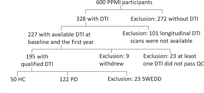
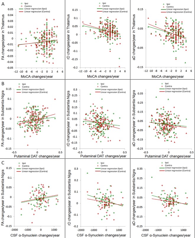
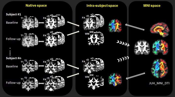
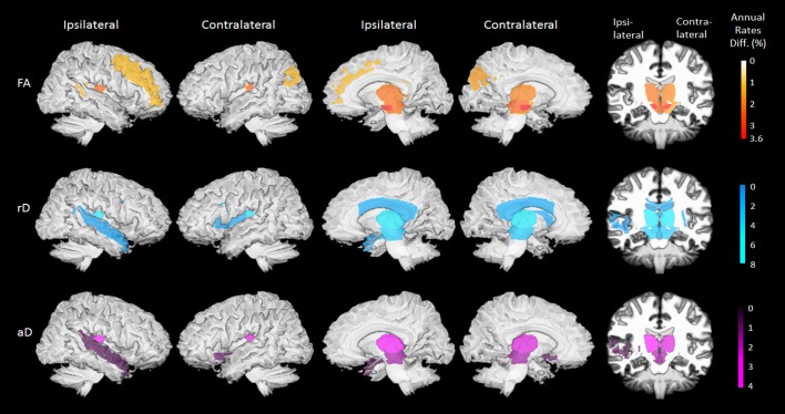
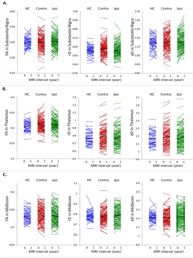
This study aimed to identify the utility of diffusion tensor imaging (DTI) in measuring the regional distribution of abnormal microstructural progression in patients with Parkinson's disease who were enrolled in the Parkinson's progression marker initiative (PPMI). One hundred and twenty two de-novo PD patients (age = 60.5±9) and 50 healthy controls (age = 60.6±11) had DTI scans at baseline and 12.6±1 months later. Automated image processing included an intra-subject registration of all time points and an inter-subjects registration to a brain atlas. Annualized rates of DTI variations including fractional anisotropy (FA), radial (rD) and axial (aD) diffusivity were estimated in a total of 118 white matter and subcortical regions of interest. A mixed effects model framework was used to determine the degree to which DTI changes differed in PD relative to changes in healthy subjects. Significant DTI changes were also tested for correlations with changes in clinical measures, dopaminergic imaging and CSF biomarkers in PD patients. Compared to normal aging, PD was associated with higher rates of FA reduction, rD and aD increases predominantly in the substantia nigra, midbrain and thalamus. The highest rates of FA reduction involved the substantia nigra (3.6±1.4%/year from baseline, whereas the highest rates of increased diffusivity involved the thalamus (rD: 8.0±2.9%/year, aD: 4.0±1.5%/year). In PD patients, high DTI changes in the substantia nigra correlated with increasing dopaminergic deficits as well as with declining α-synuclein and total tau protein concentrations in cerebrospinal fluid. Increased DTI rates in the thalamus correlated with progressive decline in global cognition in PD. The results suggest that higher rates of regional microstructural degeneration are potential markers of PD progression.
本研究旨在确定扩散张量成像(DTI)在测量参与帕金森病进展标志物计划(PPMI)的帕金森病患者异常微观结构进展的区域分布中的效用。122例初发帕金森病患者(年龄=60.5±9岁)和50例健康对照者(年龄=60.6±11岁)在基线时及12.6±1个月后进行了DTI扫描。自动化图像处理包括所有时间点的受试者内配准以及与脑图谱的受试者间配准。在总共118个白质和皮质下感兴趣区域估计了DTI变化的年化率,包括分数各向异性(FA)、径向(rD)和轴向(aD)扩散率。采用混合效应模型框架来确定帕金森病患者DTI变化与健康受试者变化相比的差异程度。还测试了帕金森病患者中显著的DTI变化与临床指标、多巴胺能成像和脑脊液生物标志物变化之间的相关性。与正常衰老相比,帕金森病主要与黑质、中脑和丘脑的FA降低率较高以及rD和aD增加率较高相关。FA降低率最高的是黑质(从基线起为3.6±1.4%/年),而扩散率增加率最高的是丘脑(rD:8.0±2.9%/年,aD:4.0±1.5%/年)。在帕金森病患者中,黑质中高DTI变化与多巴胺能缺陷增加以及脑脊液中α-突触核蛋白和总tau蛋白浓度下降相关。丘脑中DTI率增加与帕金森病患者整体认知功能的进行性下降相关。结果表明,较高的区域微观结构退变率是帕金森病进展的潜在标志物。