From the UCL Centre for Medical Imaging, University College London, 2nd Floor Charles Bell House, 43-45 Foley Street, London W1W 7TS, England (E.W.J., E.B.C., H.S.S., J.O., M.B.A., D. Atkinson, S.P.); UCL Centre for Medical Image Computing, London, England (E.B.C., U.F., B.Y., S.O., D.H., D. Alexander, E.P.); UCL Centre for Molecular Intervention, London, England (H.P., S.H., H.W.); Department of Histopathology, University College Hospital, London, England (D.P., M.R.J., A.F.); Department of Radiology (J.C.) and Centre for Medical Imaging (J.C., W.P., A.S.), University College Hospital, London, England; Division of Surgery and Interventional Science, Faculty of Medical Sciences, University College London, London, England (F.G., A.G., C.M.M., M.E.); and Department of Surgery and Cancer, Imperial College London, London, England (H.U.A.).
Radiology. 2019 May;291(2):391-397. doi: 10.1148/radiol.2019181749. Epub 2019 Apr 2.
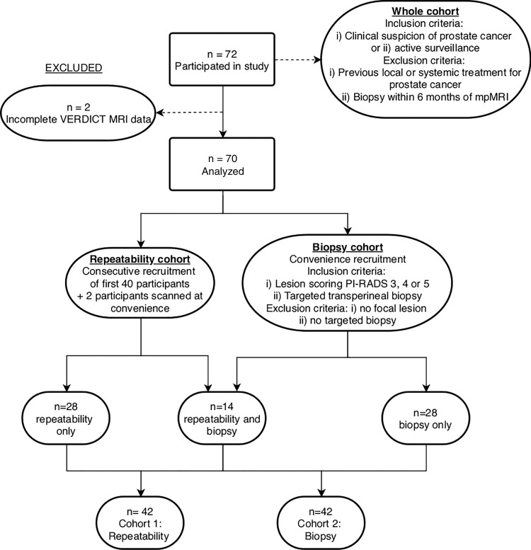
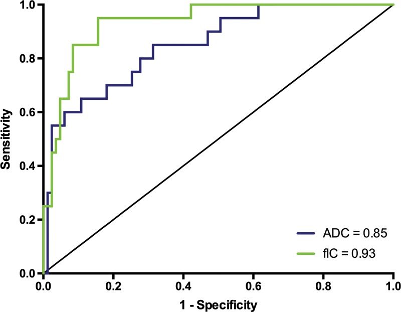
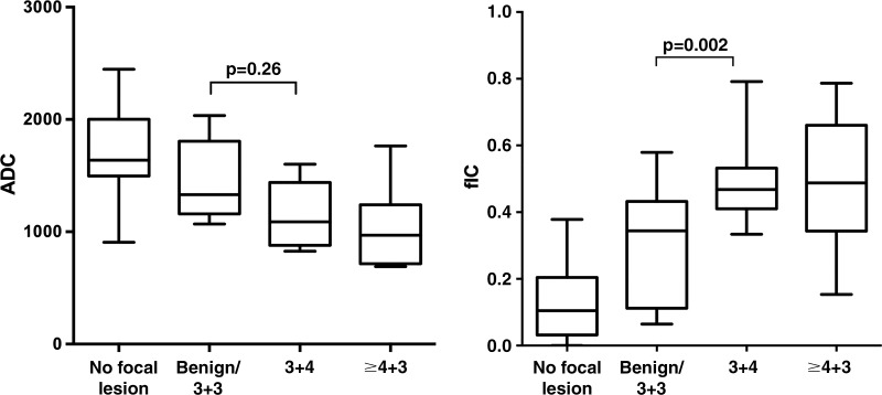
Background Biologic specificity of diffusion MRI in relation to prostate cancer aggressiveness may improve by examining separate components of the diffusion MRI signal. The Vascular, Extracellular, and Restricted Diffusion for Cytometry in Tumors (VERDICT) model estimates three distinct signal components and associates them to intracellular water, water in the extracellular extravascular space, and water in the microvasculature. Purpose To evaluate the repeatability, image quality, and diagnostic utility of intracellular volume fraction (FIC) maps obtained with VERDICT prostate MRI and to compare those maps with apparent diffusion coefficient (ADC) maps for Gleason grade differentiation. Materials and Methods Seventy men (median age, 62.2 years; range, 49.5-82.0 years) suspected of having prostate cancer or undergoing active surveillance were recruited to a prospective study between April 2016 and October 2017. All men underwent multiparametric prostate and VERDICT MRI. Forty-two of the 70 men (median age, 67.7 years; range, 50.0-82.0 years) underwent two VERDICT MRI acquisitions to assess repeatability of FIC measurements obtained with VERDICT MRI. Repeatability was measured with use of intraclass correlation coefficients (ICCs). The image quality of FIC and ADC maps was independently evaluated by two board-certified radiologists. Forty-two men (median age, 64.8 years; range, 49.5-79.6 years) underwent targeted biopsy, which enabled comparison of FIC and ADC metrics in the differentiation between Gleason grades. Results VERDICT MRI FIC demonstrated ICCs of 0.87-0.95. There was no significant difference between image quality of ADC and FIC maps (score, 3.1 vs 3.3, respectively; = .90). FIC was higher in lesions with a Gleason grade of at least 3+4 compared with benign and/or Gleason grade 3+3 lesions (mean, 0.49 ± 0.17 vs 0.31 ± 0.12, respectively; = .002). The difference in ADC between these groups did not reach statistical significance (mean, 1.42 vs 1.16 × 10 mm/sec; = .26). Conclusion Fractional intracellular volume demonstrates high repeatability and image quality and enables better differentiation of a Gleason 4 component cancer from benign and/or Gleason 3+3 histology than apparent diffusion coefficient. Published under a CC BY 4.0 license. See also the editorial by Sigmund and Rosenkrantz in this issue.
背景 扩散 MRI 的生物学特异性与前列腺癌侵袭性有关,通过检查扩散 MRI 信号的单独成分,可能会有所改善。血管、细胞外和限制扩散用于肿瘤中的细胞计数 (VERDICT) 模型估计了三个不同的信号成分,并将其与细胞内水、细胞外血管外空间中的水和微血管中的水相关联。目的 评估 VERDICT 前列腺 MRI 获得的细胞内容积分数 (FIC) 图的可重复性、图像质量和诊断效用,并比较这些图与表观扩散系数 (ADC) 图在 Gleason 分级中的差异。材料与方法 2016 年 4 月至 2017 年 10 月期间,70 名男性(中位年龄,62.2 岁;范围,49.5-82.0 岁)被招募参加一项前瞻性研究,他们怀疑患有前列腺癌或正在接受主动监测。所有男性均接受多参数前列腺和 VERDICT MRI 检查。70 名男性中有 42 名(中位年龄,67.7 岁;范围,50.0-82.0 岁)进行了两次 VERDICT MRI 采集,以评估 VERDICT MRI 获得的 FIC 测量的可重复性。使用组内相关系数 (ICC) 来衡量重复性。两名具有董事会认证的放射科医生独立评估 FIC 和 ADC 图的图像质量。42 名男性(中位年龄,64.8 岁;范围,49.5-79.6 岁)接受了靶向活检,这使我们能够比较 FIC 和 ADC 指标在区分 Gleason 分级中的作用。结果 VERDICT MRI 的 FIC 显示 ICC 为 0.87-0.95。ADC 和 FIC 图的图像质量无显著差异(评分分别为 3.1 分和 3.3 分; =.90)。与良性和/或 Gleason 3+3 病变相比,至少有 Gleason 3+4 级病变的 FIC 更高(平均值分别为 0.49 ± 0.17 和 0.31 ± 0.12; =.002)。这些组之间 ADC 的差异没有达到统计学意义(平均值分别为 1.42 和 1.16×10mm/sec; =.26)。结论 细胞内分数体积具有高重复性和图像质量,并且能够比表观扩散系数更好地区分 Gleason 4 级癌症与良性和/或 Gleason 3+3 组织学。在知识共享署名 4.0 许可下发布。请参阅本期 Sigmund 和 Rosenkrantz 的社论。