Jokivuolle Minea, Mahmood Faisal, Madsen Kristoffer Hougaard, Harbo Frederik Severin Gråe, Johnsen Lars, Lundell Henrik
Laboratory of Radiation Physics, Department of Oncology, Odense University Hospital, Odense, Denmark.
Department of Clinical Research, University of Southern Denmark, Odense, Denmark.
Med Phys. 2025 Jan;52(1):346-361. doi: 10.1002/mp.17453. Epub 2024 Oct 10.
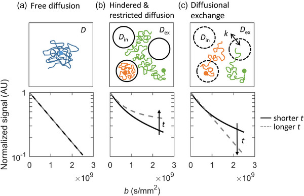
Quantitative imaging biomarkers (QIBs) can characterize tumor heterogeneity and provide information for biological guidance in radiotherapy (RT). Time-dependent diffusion MRI (TDD-MRI) derived parameters are promising QIBs, as they describe tissue microstructure with more specificity than traditional diffusion-weighted MRI (DW-MRI). Specifically, TDD-MRI can provide information about both restricted diffusion and diffusional exchange, which are the two time-dependent effects affecting diffusion in tissue, and relevant in tumors. However, exhaustive modeling of both effects can require long acquisitions and complex model fitting. Furthermore, several introduced TDD-MRI measurements can require high gradient strengths and/or complex gradient waveforms that are possibly not available in RT settings.
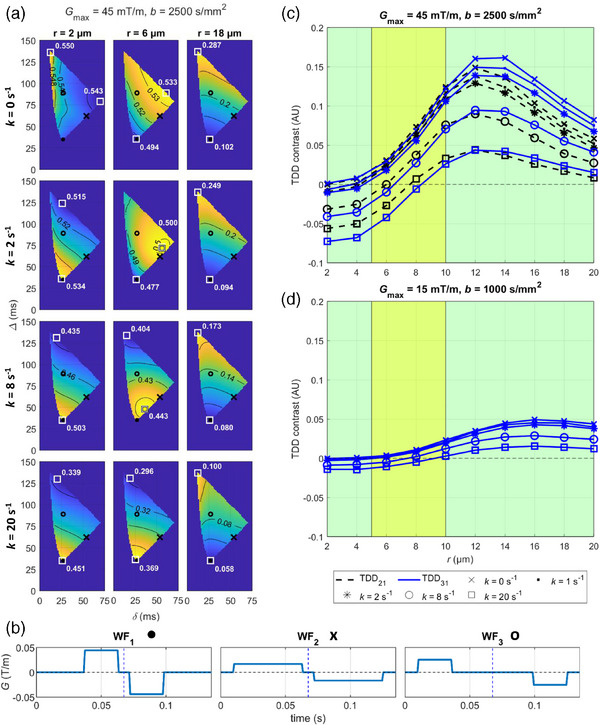
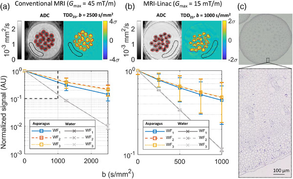
In this study, we investigated the feasibility of a simple analysis framework for the detection of restricted diffusion and diffusional exchange effects in the TDD-MRI signal. To promote the clinical applicability, we use standard gradient waveforms on a conventional 1.5 T MRI system with moderate gradient strength (G = 45 mT/m), and on a hybrid 1.5 T MRI-Linac system with low gradient strength (G = 15 mT/m).
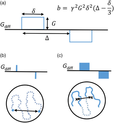
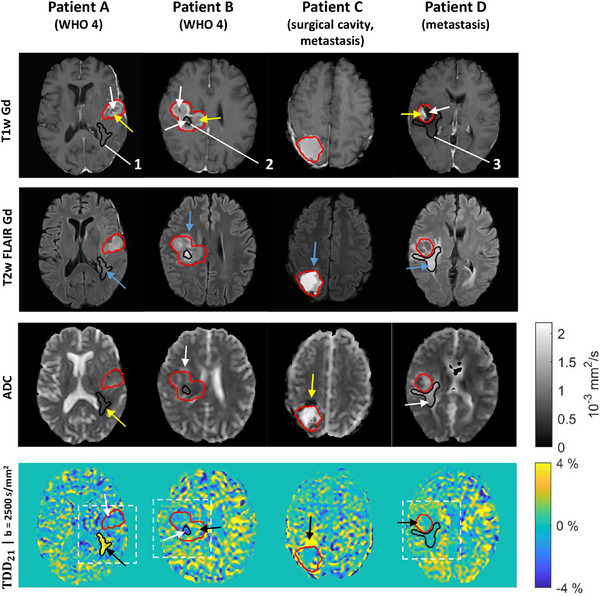
Restricted diffusion and diffusional exchange were simulated in geometries mimicking tumor microstructure to investigate the DW-MRI signal behavior and to determine optimal experimental parameters. TDD-MRI was implemented using pulsed field gradient spin echo with the optimized parameters on a conventional MRI system and a MRI-Linac. Experiments in green asparagus and 10 patients with brain lesions were performed to evaluate the time-dependent diffusion (TDD) contrast in the source DW-images.
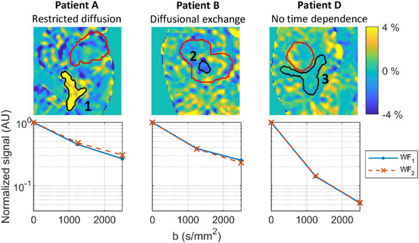
Simulations demonstrated how the TDD contrast was able to differentiate only dominating diffusional exchange in smaller cells from dominating restricted diffusion in larger cells. The maximal TDD contrast in simulations with typical cancer cell sizes and in asparagus measurements exceeded 5% on the conventional MRI but remained below 5% on the MRI-Linac. In particular, the simulated TDD contrast in typical cancer cell sizes (r = 5-10 µm) remained below or around 2% with the MRI-Linac gradient strength. In patients measured with the conventional MRI, we found sub-regions reflecting either dominating restricted diffusion or dominating diffusional exchange in and around brain lesions compared to the noisy appearing white matter.
On the conventional MRI system, the TDD contrast maps showed consistent tumor sub-regions indicating different dominating TDD effects, potentially providing information on the spatial tumor heterogeneity. On the MRI-Linac, the available TDD contrast measured in asparagus showed the same trends as with the conventional MRI but remained close to typical measurement noise levels when simulated in common cancer cell sizes. On conventional MRI systems with moderate gradient strengths, the TDD contrast could potentially be used as a tool to identify which time-dependent effects to include when choosing a biophysical model for more specific tumor characterization.
定量成像生物标志物(QIBs)可表征肿瘤异质性,并为放射治疗(RT)中的生物学指导提供信息。基于时间的扩散磁共振成像(TDD-MRI)衍生参数是很有前景的QIBs,因为它们比传统扩散加权磁共振成像(DW-MRI)更具特异性地描述组织微观结构。具体而言,TDD-MRI可提供关于受限扩散和扩散交换的信息,这是影响组织中扩散的两种随时间变化的效应,且与肿瘤相关。然而,对这两种效应进行详尽建模可能需要长时间采集和复杂的模型拟合。此外,一些引入的TDD-MRI测量可能需要高梯度强度和/或复杂的梯度波形,而这些在放疗环境中可能无法实现。
在本研究中,我们探讨了一种简单分析框架用于检测TDD-MRI信号中受限扩散和扩散交换效应的可行性。为提高临床适用性,我们在具有中等梯度强度(G = 45 mT/m)的传统1.5 T磁共振成像系统以及具有低梯度强度(G = 15 mT/m)的1.5 T磁共振成像-直线加速器混合系统上使用标准梯度波形。
在模拟肿瘤微观结构的几何模型中模拟受限扩散和扩散交换,以研究DW-MRI信号行为并确定最佳实验参数。在传统磁共振成像系统和磁共振成像-直线加速器上使用优化参数的脉冲场梯度自旋回波实现TDD-MRI。对绿芦笋和10例脑损伤患者进行实验,以评估源DW图像中的时间依赖性扩散(TDD)对比度。
模拟结果表明TDD对比度如何仅能区分较小细胞中占主导的扩散交换与较大细胞中占主导的受限扩散。在具有典型癌细胞大小的模拟以及芦笋测量中,传统磁共振成像上的最大TDD对比度超过5%,但在磁共振成像-直线加速器上仍低于5%。特别是,在磁共振成像-直线加速器梯度强度下,典型癌细胞大小(r = 5 - 10 µm)的模拟TDD对比度保持在2%以下或左右。在使用传统磁共振成像测量的患者中,与噪声明显的白质相比,我们在脑损伤内部及周围发现了反映占主导的受限扩散或占主导的扩散交换的亚区域。
在传统磁共振成像系统上,TDD对比度图显示出一致的肿瘤亚区域,表明不同的主导TDD效应,可能提供有关肿瘤空间异质性的信息。在磁共振成像-直线加速器上,在芦笋中测得的可用TDD对比度与传统磁共振成像显示出相同趋势,但在模拟常见癌细胞大小时仍接近典型测量噪声水平。在具有中等梯度强度的传统磁共振成像系统上,TDD对比度可能潜在地用作一种工具,以确定在选择用于更具体肿瘤表征的生物物理模型时应纳入哪些随时间变化的效应。