Biernacka Katarzyna B, Barańska Dobromiła, Grzelak Piotr, Czkwianianc Elżbieta, Szabelska-Zakrzewska Katarzyna
Department of Diagnostic Imagining, Polish Mother's Memorial Hospital - Research Institute, Lodz, Poland.
Department of Gastroenterology, Allergology and Paediatrics, Polish Mother's Memorial Hospital - Research Institute, Lodz, Poland.
Prz Gastroenterol. 2019;14(1):19-25. doi: 10.5114/pg.2019.83423. Epub 2019 Mar 12.
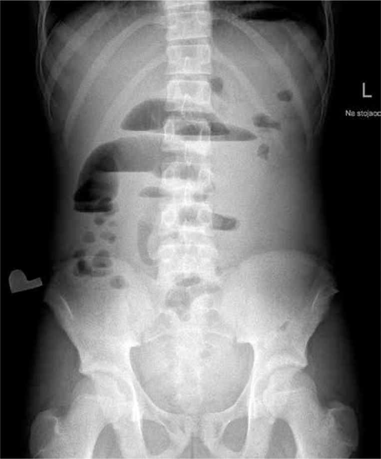
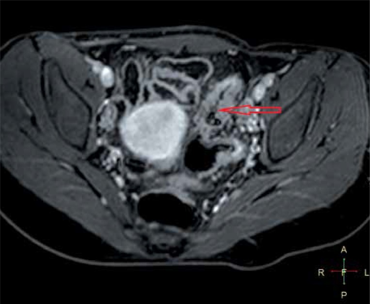
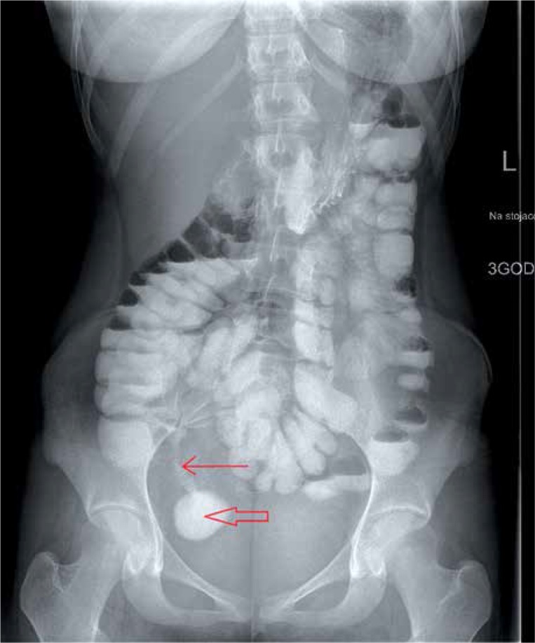
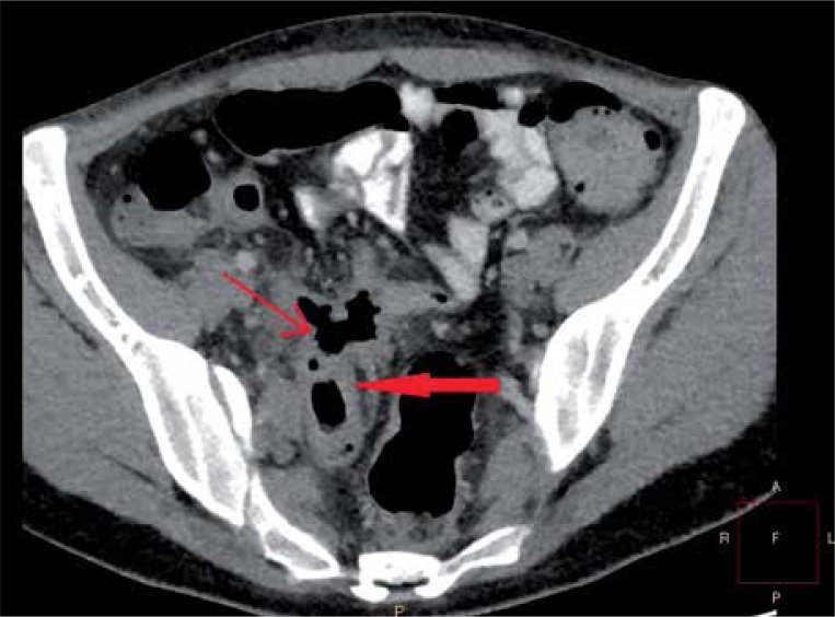
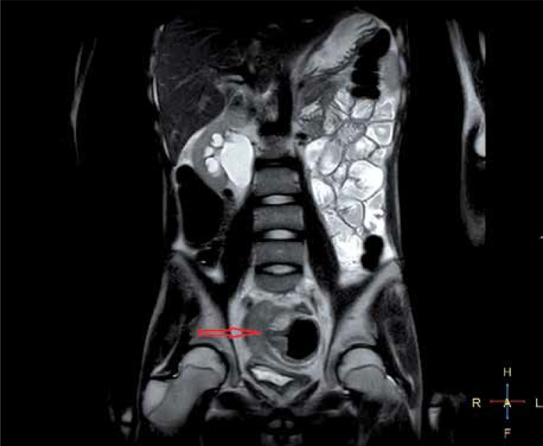
Radiological examination occupies a significant role, complementary to endoscopic studies, in the diagnostic process of inflammatory bowel disease (IBD). Both ulcerative colitis and Crohn's disease, due to multiple remissions and relapses, require repetitive examinations to evaluate the disease extent, severity, and response to pharmacological treatment. Whereas the use of barium contrast studies is progressively reduced, plain radiography confirms its utility as a first-line imaging tool for acute abdomen. Computed tomography remains an easily accessible and effective method to demonstrate disease activity and extraintestinal manifestations. However, the related radiation exposure reduces its applicability to urgent situations. Ultrasound and magnetic resonance, with the great advantage of avoiding ionising radiation, are highly recommended to present the complications of IBD. Use of oral and intravenous contrast in computed tomography enterography and magnetic resonance enterography demonstrates IBD involvement in the small intestine wall, which is difficult to assess in other radiological and endoscopic examinations.
在炎症性肠病(IBD)的诊断过程中,放射学检查是内镜检查的重要补充手段。溃疡性结肠炎和克罗恩病由于多次缓解和复发,需要反复检查以评估疾病范围、严重程度及对药物治疗的反应。尽管钡剂造影检查的使用逐渐减少,但普通X线摄影证实了其作为急腹症一线成像工具的实用性。计算机断层扫描(CT)仍然是一种易于获得且有效的方法,可用于显示疾病活动及肠外表现。然而,相关的辐射暴露限制了其在紧急情况下的应用。超声和磁共振成像(MRI)具有避免电离辐射的巨大优势,强烈推荐用于显示IBD的并发症。在CT小肠造影和MRI小肠造影中使用口服和静脉造影剂可显示IBD对小肠壁的累及情况,而这在其他放射学和内镜检查中难以评估。