Istituto di Patologia Speciale Medica, Università Cattolica del Sacro Cuore, Roma 00168, Italy.
UOC di Medicina Interna e Gastroenterologia, Area di Gastroenterologia e Oncologia Medica, Dipartimento di Scienze Gastroenterologiche, Endocrino-Metaboliche e Nefro-Urologiche, Fondazione Policlinico Universitario "A. Gemelli" IRCCS, Roma 00168, Italy.
World J Gastroenterol. 2019 Mar 28;25(12):1465-1477. doi: 10.3748/wjg.v25.i12.1465.
Anti-tumor necrosis factor α (TNFα) represents the best therapeutic option to induce mucosal healing and clinical remission in patients with moderate-severe ulcerative colitis. On the other side gut microbiota plays a crucial role in pathogenesis of ulcerative colitis but few information exists on how microbiota changes following anti-TNFα therapy and on microbiota role in mucosal healing.
To elucidate whether gut microbiota and immune system changes appear following anti TNFα therapy during dextran sulfate sodium (DSS) colitis.
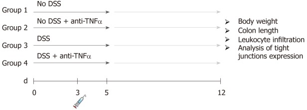
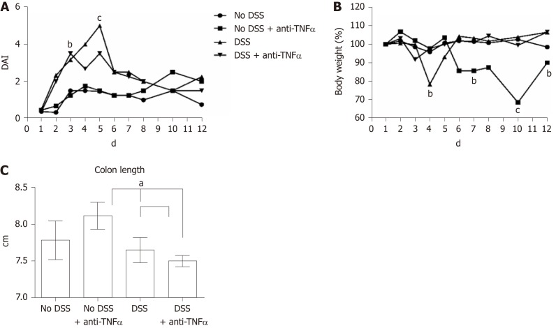
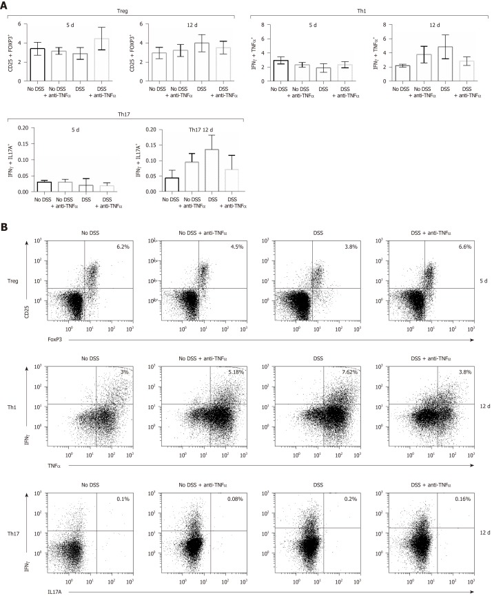
Eighty C57BL/6 mice were divided into four groups: "No DSS", "No DSS + anti-TNFα", "DSS" and "DSS + anti-TNFα". "DSS" and "DSS + anti-TNFα" were treated for 5 d with 3% DSS. At day 3, mice whithin "No DSS+anti-TNFα" and "DSS+anti-TNFα" group received 5 mg/kg of an anti-TNFα agent. Forty mice were sacrificed at day 5, forty at day 12, after one week of recovery post DSS. The severity of colitis was assessed by a clinical score (Disease Activity Index), colon length and histology. Bacteria such as , , and () were evaluated by quantitative PCR. Type 1 helper T lymphocytes (Th1), type 17 helper T lymphocytes (Th17) and CD4 regulatory T lymphocytes (Treg) distributions in the mesenteric lymph node (MLN) were studied by flow cytometry.
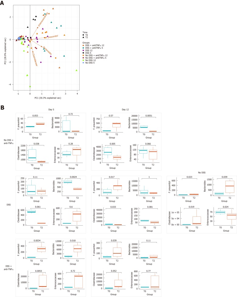
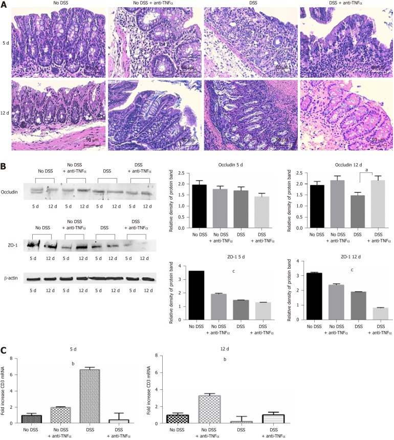
Bacteria associated with a healthy state (., such as , and ) decreased during colitis and increased in course of anti-TNFα treatment. Conversely, microorganisms belonging to genera, which are linked to inflammatory processes, showed an opposite trend. Furthermore, in colitic mice treated with anti-TNFα microbial changes were associated with an initial increase (day 5 of the colitis) in Treg cells and a consequent decrease (day 12 post DSS) in Th1 and Th17 frequency cells. Healthy mice treated with anti-TNFα showed the same histological, microbial and immune features of untreated colitic mice. "No DSS + anti-TNFα" group showed a lymphomononuclear infiltrate both at 5 and 12 d at hematoxylin and eosin staining, an increase of in Th1 and Th17 frequency at day 12, an increase of at day 5, a decrease of and at day 12.
Anti-TNFα treatment in experimental model of colitis improves disease activity but it is associated to an increase in Th17 pathway together with gut microbiota alteration.
抗肿瘤坏死因子 α(TNFα)是诱导中重度溃疡性结肠炎黏膜愈合和临床缓解的最佳治疗选择。另一方面,肠道微生物群在溃疡性结肠炎的发病机制中起着至关重要的作用,但关于抗 TNFα 治疗后微生物群的变化以及微生物群在黏膜愈合中的作用的信息很少。
阐明在葡聚糖硫酸钠(DSS)结肠炎期间,抗 TNFα 治疗后肠道微生物群和免疫系统是否发生变化。
将 80 只 C57BL/6 小鼠分为四组:“无 DSS”、“无 DSS+抗 TNFα”、“DSS”和“DSS+抗 TNFα”。“DSS”和“DSS+抗 TNFα”用 3% DSS 处理 5 天。在第 3 天,“无 DSS+抗 TNFα”和“DSS+抗 TNFα”组的小鼠接受 5mg/kg 的抗 TNFα 药物。40 只小鼠在第 5 天,40 只在第 12 天(DSS 后 1 周恢复后)处死。通过临床评分(疾病活动指数)、结肠长度和组织学评估结肠炎的严重程度。通过定量 PCR 评估细菌,如 、 、 ()。通过流式细胞术研究肠系膜淋巴结(MLN)中 1 型辅助 T 淋巴细胞(Th1)、17 型辅助 T 淋巴细胞(Th17)和 CD4 调节性 T 淋巴细胞(Treg)的分布。
在结肠炎期间,与健康状态相关的细菌(例如 、 、 )减少,并在抗 TNFα 治疗过程中增加。相反,属于 属的与炎症过程相关的微生物表现出相反的趋势。此外,在接受抗 TNFα 治疗的结肠炎小鼠中,微生物变化与初始 Treg 细胞增加(结肠炎第 5 天)和随后 Th1 和 Th17 频率细胞减少(DSS 后第 12 天)相关。接受抗 TNFα 治疗的健康小鼠表现出与未经治疗的结肠炎小鼠相同的组织学、微生物和免疫特征。“无 DSS+抗 TNFα”组在第 5 天和第 12 天的苏木精和伊红染色中均显示出淋巴单核细胞浸润,第 12 天 Th1 和 Th17 频率增加,第 5 天 增加,第 12 天 和 减少。
在结肠炎的实验模型中,抗 TNFα 治疗可改善疾病活动度,但与 Th17 途径的增加以及肠道微生物群的改变相关。