Oxford Parkinson's Disease Centre, Department of Physiology, Anatomy and Genetics, University of Oxford, Oxford, UK.
Oxford Parkinson's Disease Centre, Department of Physiology, Anatomy and Genetics, University of Oxford, Oxford, UK.
Neurobiol Dis. 2019 Jul;127:512-526. doi: 10.1016/j.nbd.2019.04.005. Epub 2019 Apr 5.
Mutations in LRRK2 are the most common cause of autosomal dominant Parkinson's disease, and the relevance of LRRK2 to the sporadic form of the disease is becoming ever more apparent. It is therefore essential that studies are conducted to improve our understanding of the cellular role of this protein. Here we use multiple models and techniques to identify the pathways through which LRRK2 mutations may lead to the development of Parkinson's disease.
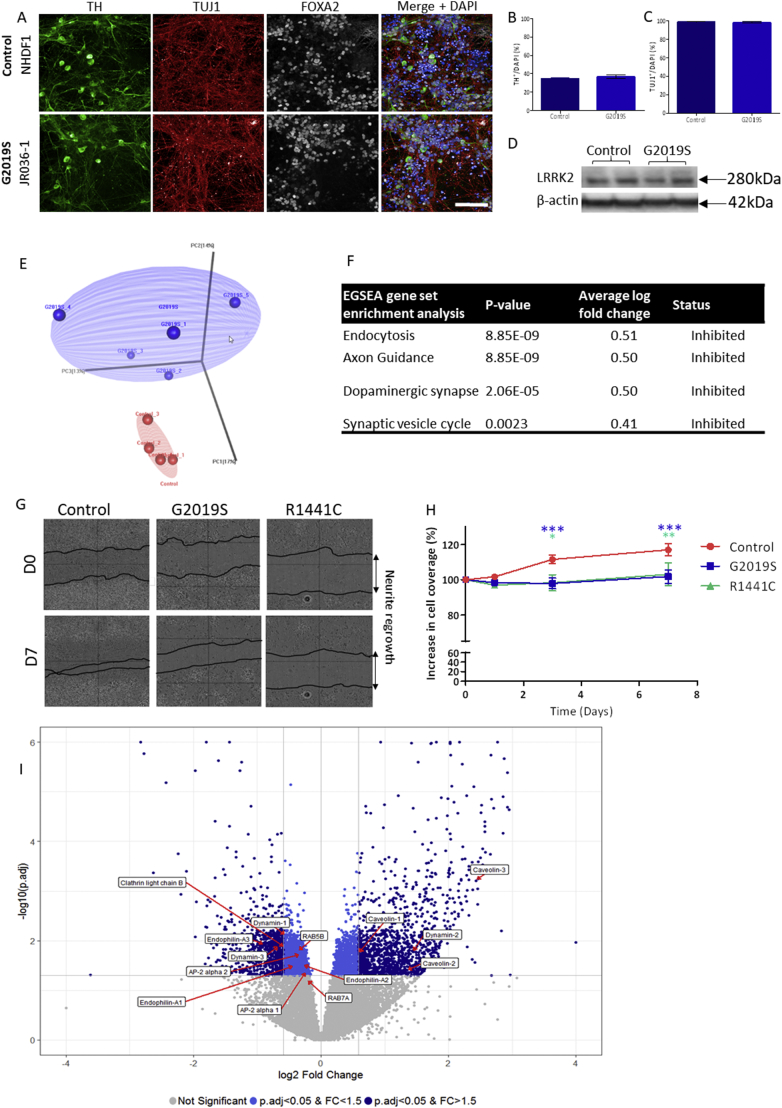
A novel integrated transcriptomics and proteomics approach was used to identify pathways that were significantly altered in iPSC-derived dopaminergic neurons carrying the LRRK2-G2019S mutation. Western blotting, immunostaining and functional assays including FM1-43 analysis of synaptic vesicle endocytosis were performed to confirm these findings in iPSC-derived dopaminergic neuronal cultures carrying either the LRRK2-G2019S or the LRRK2-R1441C mutation, and LRRK2 BAC transgenic rats, and post-mortem human brain tissue from LRRK2-G2019S patients.
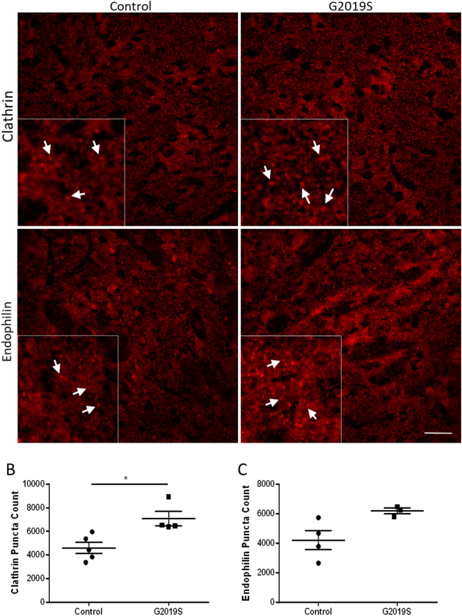
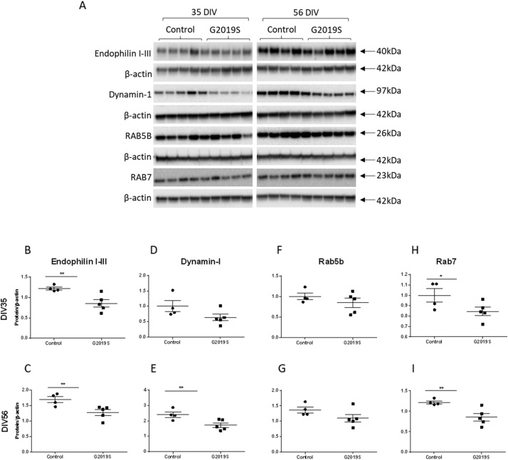
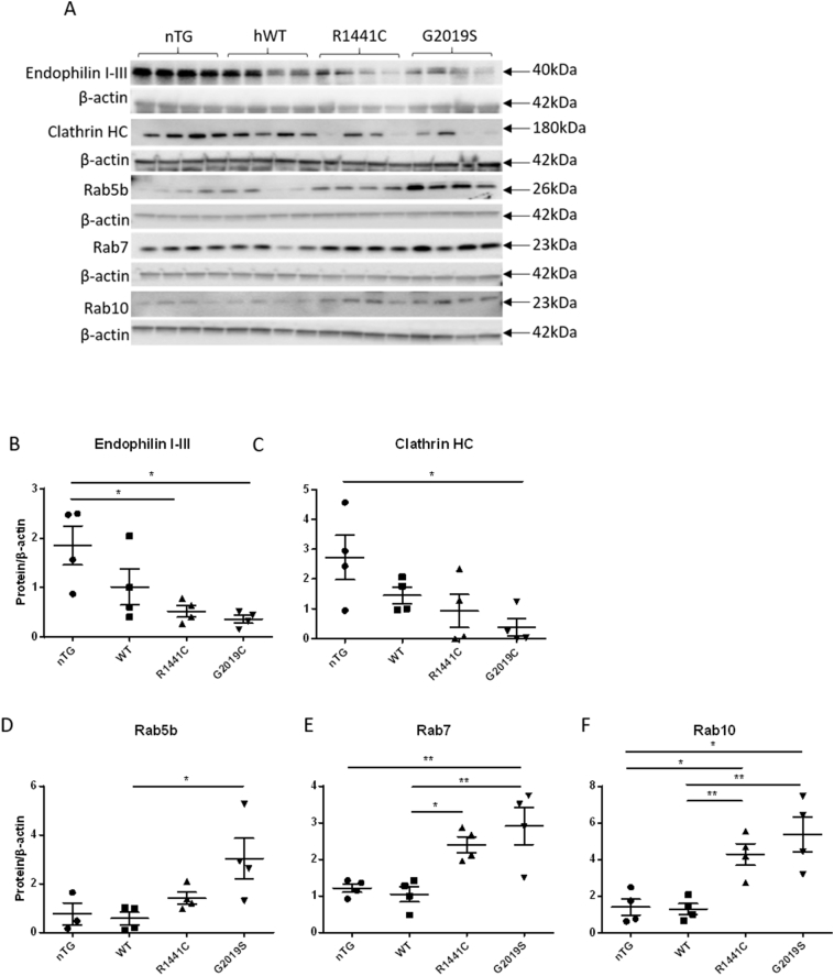
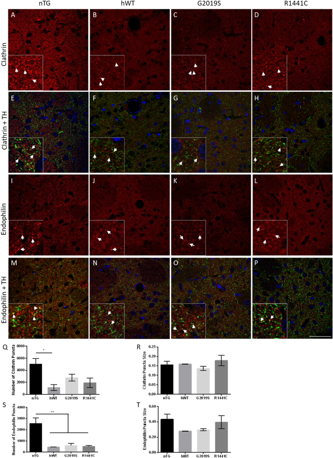
Our integrated -omics analysis revealed highly significant dysregulation of the endocytic pathway in iPSC-derived dopaminergic neurons carrying the LRRK2-G2019S mutation. Western blot analysis confirmed that key endocytic proteins including endophilin I-III, dynamin-1, and various RAB proteins were downregulated in these cultures and in cultures carrying the LRRK2-R1441C mutation, compared with controls. We also found changes in expression of 25 RAB proteins. Changes in endocytic protein expression led to a functional impairment in clathrin-mediated synaptic vesicle endocytosis. Further to this, we found that the endocytic pathway was also perturbed in striatal tissue of aged LRRK2 BAC transgenic rats overexpressing either the LRRK2 wildtype, LRRK2-R1441C or LRRK2-G2019S transgenes. Finally, we found that clathrin heavy chain and endophilin I-III levels are increased in human post-mortem tissue from LRRK2-G2019S patients compared with controls.
Our study demonstrates extensive alterations across the endocytic pathway associated with LRRK2 mutations in iPSC-derived dopaminergic neurons and BAC transgenic rats, as well as in post-mortem brain tissue from PD patients carrying a LRRK2 mutation. In particular, we find evidence of disrupted clathrin-mediated endocytosis and suggest that LRRK2-mediated PD pathogenesis may arise through dysregulation of this process.
LRRK2 突变是常染色体显性遗传帕金森病最常见的病因,LRRK2 与散发性帕金森病的相关性越来越明显。因此,进行研究以提高我们对该蛋白细胞功能的理解是至关重要的。在这里,我们使用多种模型和技术来确定 LRRK2 突变可能导致帕金森病发展的途径。
使用一种新的整合转录组学和蛋白质组学方法来鉴定在携带 LRRK2-G2019S 突变的 iPSC 衍生的多巴胺能神经元中显著改变的途径。进行 Western blot 分析、免疫染色和功能测定,包括 FM1-43 分析突触小泡内吞作用,以确认在携带 LRRK2-G2019S 或 LRRK2-R1441C 突变的 iPSC 衍生的多巴胺能神经元培养物以及 LRRK2 BAC 转基因大鼠和携带 LRRK2-G2019S 突变的人类死后脑组织中发现这些结果。
我们的整合组学分析显示,在携带 LRRK2-G2019S 突变的 iPSC 衍生的多巴胺能神经元中,内吞途径高度失调。Western blot 分析证实,与对照组相比,这些培养物和携带 LRRK2-R1441C 突变的培养物中关键的内吞蛋白,包括内吞素 I-III、动力蛋白-1 和各种 RAB 蛋白下调。我们还发现 25 种 RAB 蛋白的表达发生变化。内吞蛋白表达的变化导致网格蛋白介导的突触小泡内吞作用的功能障碍。此外,我们发现,在过表达野生型 LRRK2、LRRK2-R1441C 或 LRRK2-G2019S 转基因的老年 LRRK2 BAC 转基因大鼠的纹状体组织中,内吞途径也受到干扰。最后,我们发现与对照组相比,LRRK2-G2019S 患者的人死后脑组织中的网格蛋白重链和内吞素 I-III 水平增加。
我们的研究表明,在 iPSC 衍生的多巴胺能神经元和 BAC 转基因大鼠以及携带 LRRK2 突变的 PD 患者的死后脑组织中,与 LRRK2 突变相关的内吞途径广泛改变。特别是,我们发现有证据表明网格蛋白介导的内吞作用受到破坏,并表明 LRRK2 介导的 PD 发病机制可能是通过该过程的失调而产生的。