Erciyes University Medical Faculty, Department of Radiology, Kayseri, Turkey.
Erciyes University Medical Faculty, Department of Otorhinolaryngology, Kayseri, Turkey.
Braz J Otorhinolaryngol. 2020 Jul-Aug;86(4):468-482. doi: 10.1016/j.bjorl.2019.02.003. Epub 2019 Mar 16.
Squamous cell carcinoma is the most common laryngeal neoplasm and accounts for approximately 95% of all malignant neoplams of the larynx. However, various benign and malignant tumors and inflammatory diseases may affect the larynx.
The purpose of this study is to analyze the clinical and imaging findings of non-squamous cell neoplasms and inflammatory diseases of the larynx.
This retrospective study was conducted in 18 patients who were diagnosed with non-squamous cell carcinoma lesions of larynx at our institution between 2007-2017. Clinical symptoms, examination findings, imaging characteristics, histopathologic diagnosis and treatment modalities were analyzed.
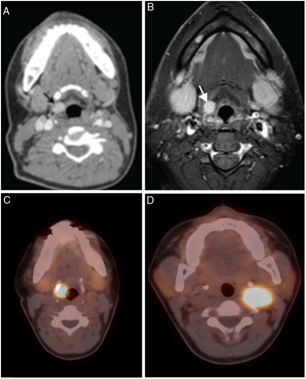
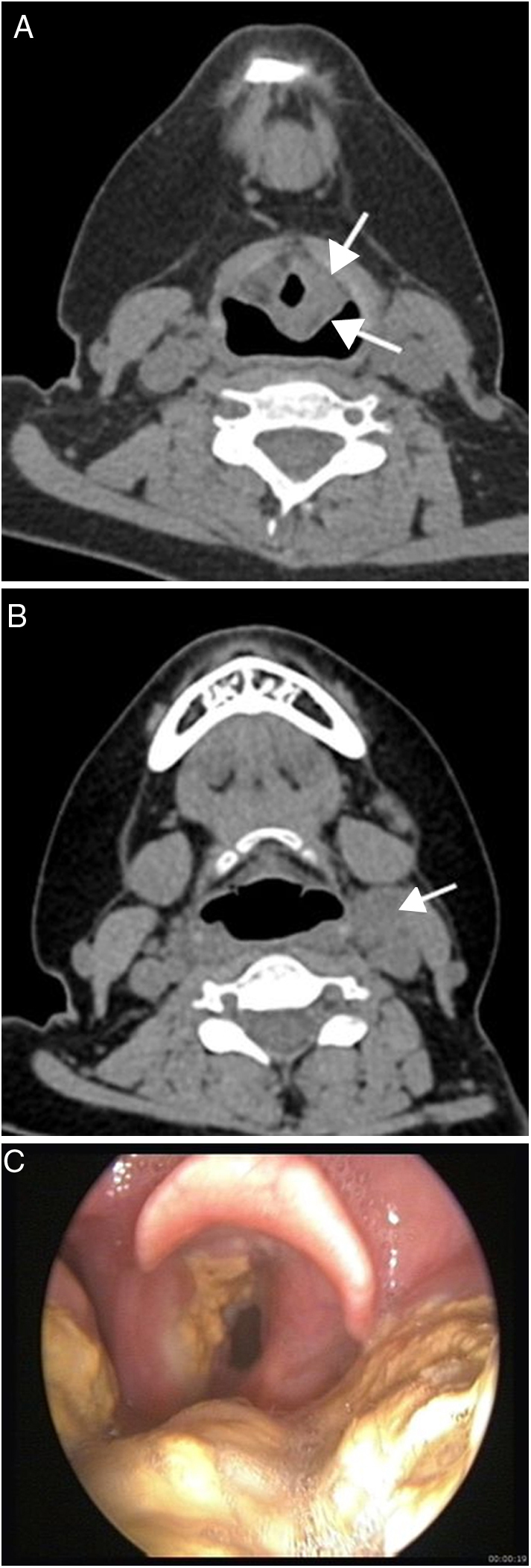
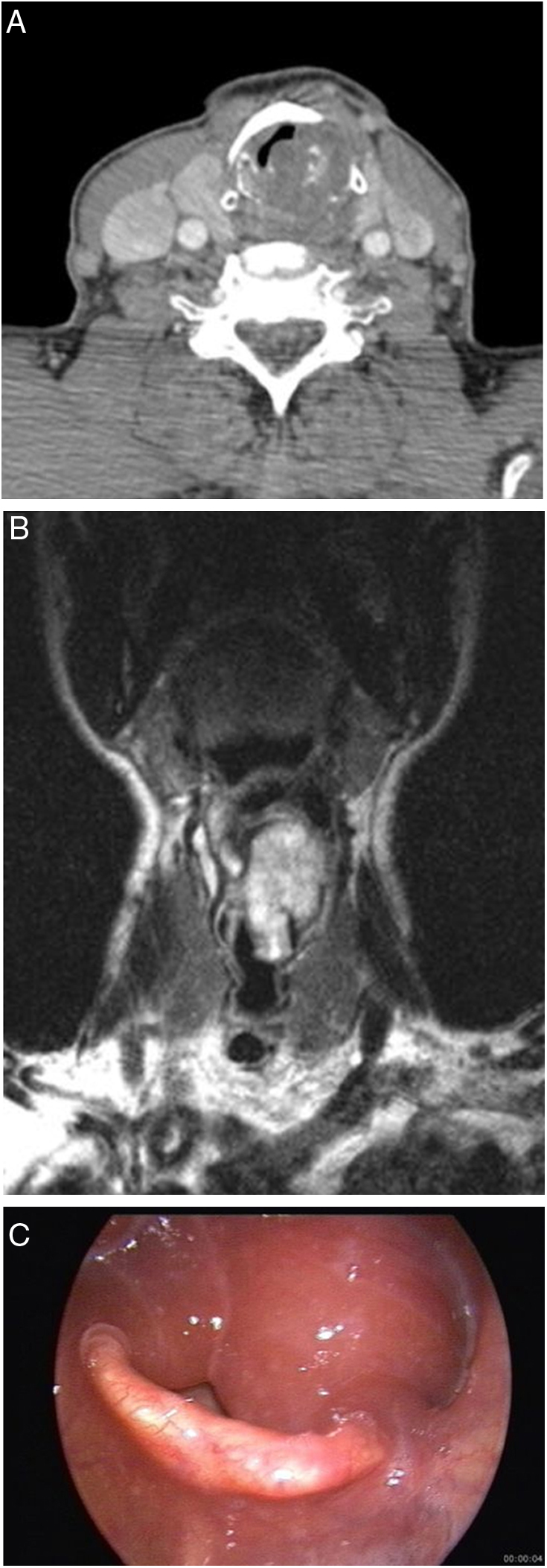
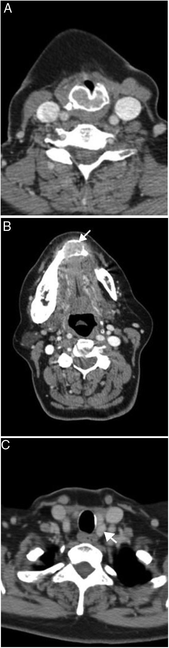
There were 9 malignant lesions (2 chondrosarcoma, 1 neuroendocrine tumor-atipical carcinoid, 1 Natural Killer/T-cell lymphoma, 1 diffuse large B-cell lymphoma, 3 plasmocytoma-multiple myeloma involvement, 1 adenocarcinoma metastasis), 3 benign neoplasms (chondroma, paraganglioma, lipoma), 2 tumor-like lesions (Brown tumor and inflammatory myofibroblastic tumor), 3 inflammatory lesions (Wegener granulomatosis, Behçet's disease and tuberculosis involvements), and 1 vascular malformation. The most common presenting symptom was hoarseness (66.6%). Paraganglioma was seen as hypervascular lesion on computed tomography and magnetic resonance imaging and showed intense tracer uptake on 68Gallium-DOTA-peptide PET/CT. Chondroid matrix calcifications were detected in chondroma and chondrosarcoma-grade 1. In patients with vascular malformation and lipoma, the typical imaging findings made it possible to diagnose.
Imaging studies may provide clues for diagnosis of non-squamous cell laryngeal lesions. Clinical and imaging findings and previous clinical history should be evaluated together in clinical management of laryngeal lesions.
鳞状细胞癌是最常见的喉部肿瘤,约占喉部所有恶性肿瘤的 95%。然而,各种良性和恶性肿瘤以及炎症性疾病也可能影响喉部。
本研究旨在分析喉部非鳞状细胞肿瘤和炎症性疾病的临床和影像学表现。
本回顾性研究纳入了 2007 年至 2017 年间在我院诊断为喉部非鳞状细胞癌病变的 18 例患者。分析了临床症状、检查结果、影像学特征、组织病理学诊断和治疗方式。
有 9 例恶性病变(2 例软骨肉瘤、1 例神经内分泌肿瘤-不典型类癌、1 例自然杀伤/T 细胞淋巴瘤、1 例弥漫性大 B 细胞淋巴瘤、3 例浆细胞瘤-多发性骨髓瘤累及、1 例腺癌转移)、3 例良性肿瘤(软骨瘤、副神经节瘤、脂肪瘤)、2 例肿瘤样病变(棕色瘤和炎症性肌纤维母细胞瘤)、3 例炎症性病变(韦格纳肉芽肿、贝切特病和结核累及)和 1 例血管畸形。最常见的表现症状是声音嘶哑(66.6%)。副神经节瘤在 CT 和磁共振成像上表现为富血管病变,68Ga-DOTA-肽 PET/CT 显示强烈的示踪剂摄取。软骨瘤和 1 级软骨肉瘤中检测到软骨样基质钙化。对于血管畸形和脂肪瘤患者,典型的影像学表现有助于诊断。
影像学研究可为喉部非鳞状细胞病变的诊断提供线索。在喉部病变的临床管理中,应综合评估临床和影像学表现以及既往临床病史。