• 文献检索
  • 文档翻译
  • 深度研究
  • 学术资讯
  • Suppr Zotero 插件Zotero 插件
  • 邀请有礼
  • 套餐&价格
  • 历史记录
应用&插件
Suppr Zotero 插件Zotero 插件浏览器插件Mac 客户端Windows 客户端微信小程序
定价
高级版会员购买积分包购买API积分包
服务
文献检索文档翻译深度研究API 文档MCP 服务
关于我们
关于 Suppr公司介绍联系我们用户协议隐私条款
关注我们

Suppr 超能文献

核心技术专利:CN118964589B侵权必究
粤ICP备2023148730 号-1Suppr @ 2026

文献检索

告别复杂PubMed语法,用中文像聊天一样搜索,搜遍4000万医学文献。AI智能推荐,让科研检索更轻松。

立即免费搜索

文件翻译

保留排版,准确专业,支持PDF/Word/PPT等文件格式,支持 12+语言互译。

免费翻译文档

深度研究

AI帮你快速写综述,25分钟生成高质量综述,智能提取关键信息,辅助科研写作。

立即免费体验

超声造影时间-强度曲线分析在喉鳞状细胞癌非骨化性甲状腺软骨侵犯中的应用

Time-Intensity Curve Analysis of Contrast-Enhanced Ultrasound for Non-Ossified Thyroid Cartilage Invasion in Laryngeal Squamous Cell Carcinoma.

作者信息

Pucėtaitė Milda, Mitraitė Dalia, Tarasevičius Rytis, Farina Davide, Ryškienė Silvija, Lukoševičius Saulius, Padervinskis Evaldas, Šarauskas Valdas, Vaitkus Saulius

机构信息

Department of Radiology, Faculty of Medicine, Medical Academy, Lithuanian University of Health Sciences, A. Mickevičiaus Str. 9, 44307 Kaunas, Lithuania.

Department of Radiology, Lithuanian University of Health Sciences Kaunas Clinics, Eivenių 2, 50009 Kaunas, Lithuania.

出版信息

Tomography. 2025 May 16;11(5):57. doi: 10.3390/tomography11050057.

DOI:10.3390/tomography11050057
PMID:40423259
原文链接:https://pmc.ncbi.nlm.nih.gov/articles/PMC12115638/
Abstract

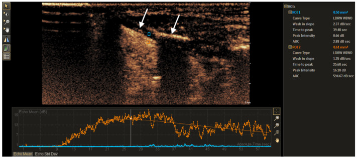
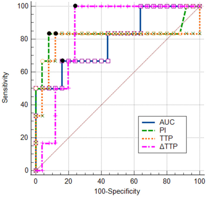
This study aimed to assess the diagnostic value of contrast-enhanced ultrasound (CEUS) time-intensity curve (TIC) parameters in detecting non-ossified thyroid cartilage invasion in patients with laryngeal squamous cell carcinoma (SCC). : A CEUS TIC analysis was performed on 32 cases from 27 patients with histologically confirmed laryngeal SCC. The diagnostic performance of time to peak (TTP), peak intensity (PI), wash-in slope (WIS), area under the curve (AUC), and their quantitative differences (∆TTP, ∆PI, ∆WIS, and ∆AUC) to discriminate between the invaded and the non-invaded non-ossified thyroid cartilage was determined using ROC analysis. A logistic regression analysis was employed to identify significant predictors. : In an ROC analysis, of all TIC parameters analyzed separately, ∆TTP showed the greatest diagnostic performance (AUC: 0.85). A ∆TTP cut-off of ≤ 8.9 s differentiated between the invaded and the non-invaded non-ossified thyroid cartilage with a sensitivity of 100%, specificity of 76.9%, and accuracy of 81.3%. A combination of ∆TTP and PI increased the AUC to 0.93, specificity to 100%, and accuracy to 96.8%, but reduced the sensitivity to 83.3%. Meanwhile, the visual assessment of enhancement on CEUS to detect cartilage invasion had 83.3% sensitivity and 84.6% specificity. In a univariate logistic regression, only ∆TTP was a significant predictor of non-ossified thyroid cartilage invasion (OR: 0.80; 95% CI: 0.64-1.00). For every second increase in ∆TTP, the probability of thyroid cartilage invasion decreased by 20%. : CEUS TIC parameters, particularly a combination of ∆TTP and PI, showed high diagnostic performance in the detection of non-ossified thyroid cartilage invasion in laryngeal SCC.

摘要

本研究旨在评估超声造影(CEUS)时间-强度曲线(TIC)参数在检测喉鳞状细胞癌(SCC)患者非骨化甲状腺软骨侵犯中的诊断价值。对27例经组织学证实为喉SCC的患者的32个病例进行了CEUS TIC分析。采用ROC分析确定达峰时间(TTP)、峰值强度(PI)、流入斜率(WIS)、曲线下面积(AUC)及其定量差异(∆TTP、∆PI、∆WIS和∆AUC)在鉴别侵袭性和非侵袭性非骨化甲状腺软骨方面的诊断性能。采用逻辑回归分析确定显著预测因素。在ROC分析中,单独分析的所有TIC参数中,∆TTP显示出最大的诊断性能(AUC:0.85)。∆TTP截断值≤8.9 s可区分侵袭性和非侵袭性非骨化甲状腺软骨,敏感性为100%,特异性为76.9%,准确性为81.3%。∆TTP和PI联合可将AUC提高到0.93,特异性提高到100%,准确性提高到96.8%,但敏感性降低到83.3%。同时,CEUS增强的视觉评估检测软骨侵犯的敏感性为83.3%,特异性为84.6%。在单因素逻辑回归中,只有∆TTP是非骨化甲状腺软骨侵犯的显著预测因素(OR:0.80;95%CI:0.64-1.00)。∆TTP每增加1秒,甲状腺软骨侵犯的概率降低20%。CEUS TIC参数,特别是∆TTP和PI联合,在检测喉SCC非骨化甲状腺软骨侵犯方面显示出较高的诊断性能。

https://cdn.ncbi.nlm.nih.gov/pmc/blobs/2237/12115638/194f0e56dc35/tomography-11-00057-g004.jpg
https://cdn.ncbi.nlm.nih.gov/pmc/blobs/2237/12115638/82b19227c4de/tomography-11-00057-g001.jpg
https://cdn.ncbi.nlm.nih.gov/pmc/blobs/2237/12115638/a34bad3aec69/tomography-11-00057-g002.jpg
https://cdn.ncbi.nlm.nih.gov/pmc/blobs/2237/12115638/773e76fc805c/tomography-11-00057-g003.jpg
https://cdn.ncbi.nlm.nih.gov/pmc/blobs/2237/12115638/194f0e56dc35/tomography-11-00057-g004.jpg

相似文献

1
Time-Intensity Curve Analysis of Contrast-Enhanced Ultrasound for Non-Ossified Thyroid Cartilage Invasion in Laryngeal Squamous Cell Carcinoma.超声造影时间-强度曲线分析在喉鳞状细胞癌非骨化性甲状腺软骨侵犯中的应用
Tomography. 2025 May 16;11(5):57. doi: 10.3390/tomography11050057.
2
CT-scan prediction of thyroid cartilage invasion for early laryngeal squamous cell carcinoma.CT 扫描预测早期喉鳞癌侵犯甲状软骨。
Eur Arch Otorhinolaryngol. 2013 Jan;270(1):287-91. doi: 10.1007/s00405-012-2005-8. Epub 2012 Mar 30.
3
CT-based radiomics features in the prediction of thyroid cartilage invasion from laryngeal and hypopharyngeal squamous cell carcinoma.基于 CT 的放射组学特征预测喉及下咽鳞状细胞癌侵犯甲状软骨。
Cancer Imaging. 2020 Nov 11;20(1):81. doi: 10.1186/s40644-020-00359-2.
4
The Diagnostic Value of CEUS in Assessing Non-Ossified Thyroid Cartilage Invasion in Patients with Laryngeal Squamous Cell Carcinoma.超声造影在评估喉鳞状细胞癌患者非骨化性甲状软骨侵犯中的诊断价值
J Clin Med. 2024 Feb 3;13(3):891. doi: 10.3390/jcm13030891.
5
Comparison of MR Imaging and Dual-Energy CT for the Evaluation of Cartilage Invasion by Laryngeal and Hypopharyngeal Squamous Cell Carcinoma.磁共振成像与双能量 CT 对喉及下咽鳞状细胞癌软骨侵犯评估的比较。
AJNR Am J Neuroradiol. 2018 Mar;39(3):524-531. doi: 10.3174/ajnr.A5530. Epub 2018 Jan 25.
6
Is there a role for PET/CT parameters to differentiate thyroid cartilage invasion from penetration?PET/CT参数在区分甲状腺软骨侵犯与穿透方面是否有作用?
Eur J Radiol. 2016 Feb;85(2):319-23. doi: 10.1016/j.ejrad.2015.11.039. Epub 2015 Dec 7.
7
Thyroid cartilage invasion in laryngeal and hypopharyngeal squamous cell carcinoma treated with total laryngectomy.全喉切除术治疗的喉及下咽鳞状细胞癌中的甲状腺软骨侵犯
Eur Arch Otorhinolaryngol. 2016 Nov;273(11):3789-3794. doi: 10.1007/s00405-016-4120-4. Epub 2016 Jun 2.
8
Pretherapeutic staging of hypopharyngeal carcinoma. Clinical findings, computed tomography, and magnetic resonance imaging compared with histopathologic evaluation.下咽癌的治疗前分期。将临床检查结果、计算机断层扫描和磁共振成像与组织病理学评估进行比较。
Arch Otolaryngol Head Neck Surg. 1997 Sep;123(9):908-13. doi: 10.1001/archotol.1997.01900090016003.
9
Effectiveness of ultrasonography and computed tomography in assessing thyroid cartilage invasion in laryngeal and hypopharyngeal cancers.超声检查和计算机断层扫描在评估喉癌和下咽癌中甲状腺软骨侵犯情况的有效性
J Ultrasound. 2017 Aug 19;20(3):205-211. doi: 10.1007/s40477-017-0259-0. eCollection 2017 Sep.
10
[Diagnostic value of enhanced CT/MRI for thyroid cartilage invasion by malignant tumor].[增强CT/MRI对甲状腺软骨受恶性肿瘤侵犯的诊断价值]
Zhonghua Er Bi Yan Hou Tou Jing Wai Ke Za Zhi. 2017 May 7;52(5):372-376. doi: 10.3760/cma.j.issn.1673-0860.2017.05.010.

本文引用的文献

1
The Diagnostic Value of CEUS in Assessing Non-Ossified Thyroid Cartilage Invasion in Patients with Laryngeal Squamous Cell Carcinoma.超声造影在评估喉鳞状细胞癌患者非骨化性甲状软骨侵犯中的诊断价值
J Clin Med. 2024 Feb 3;13(3):891. doi: 10.3390/jcm13030891.
2
Quantitative time intensity curve analysis of contrast-enhanced ultrasound (CEUS) examinations for the assessment of focal liver lesions.超声造影定量时间强度曲线分析在局灶性肝脏病变评估中的应用。
Med Ultrason. 2024 Mar 27;26(1):63-71. doi: 10.11152/mu-4108. Epub 2023 Nov 6.
3
Concordance between head and neck MRI and histopathology in detecting laryngeal subsite invasion among patients with laryngeal cancer.
头颈部 MRI 与组织病理学在检测喉癌患者喉内不同亚部位侵犯的一致性。
Cancer Imaging. 2023 Oct 19;23(1):99. doi: 10.1186/s40644-023-00618-y.
4
The clinical value of DCE-MRI for differentiating secondary laryngeal cartilage lesions.DCE-MRI 对鉴别继发性喉软骨病变的临床价值。
Medicine (Baltimore). 2023 Mar 31;102(13):e33352. doi: 10.1097/MD.0000000000033352.
5
Salvage vs. Primary Total Laryngectomy in Patients with Locally Advanced Laryngeal or Hypopharyngeal Carcinoma: Oncologic Outcomes and Their Predictive Factors.局部晚期喉癌或下咽癌患者挽救性手术与初次全喉切除术的疗效及预测因素分析
J Clin Med. 2023 Feb 7;12(4):1305. doi: 10.3390/jcm12041305.
6
Normalized Parameters of Dynamic Contrast-Enhanced Perfusion MRI and DWI-ADC for Differentiation between Posttreatment Changes and Recurrence in Head and Neck Cancer.动态对比增强磁共振灌注成像和 DWI-ADC 标准化参数在头颈部癌症治疗后改变与复发鉴别中的应用。
AJNR Am J Neuroradiol. 2022 Aug;43(8):1184-1189. doi: 10.3174/ajnr.A7567. Epub 2022 Jul 14.
7
Contrast-Enhanced Ultrasonography for Differential Diagnosis of Benign and Malignant Thyroid Lesions: Single-Institutional Prospective Study of Qualitative and Quantitative CEUS Characteristics.超声造影在甲状腺良恶性病变鉴别诊断中的应用:单中心前瞻性研究定性及定量 CEUS 特征。
Contrast Media Mol Imaging. 2022 Apr 23;2022:8229445. doi: 10.1155/2022/8229445. eCollection 2022.
8
Imaging features of cartilaginous tumors of the head and neck.头颈部软骨肿瘤的影像学特征。
J Clin Imaging Sci. 2021 Dec 4;11:66. doi: 10.25259/JCIS_186_2021. eCollection 2021.
9
MR Imaging of Laryngeal and Hypopharyngeal Cancer.喉和下咽癌的磁共振成像。
Magn Reson Imaging Clin N Am. 2022 Feb;30(1):53-72. doi: 10.1016/j.mric.2021.08.002.
10
Qualitative and Quantitative Contrast-Enhanced Ultrasound Combined with Conventional Ultrasound for Predicting the Malignancy of Soft Tissue Tumors.定性和定量对比增强超声联合常规超声预测软组织肿瘤的恶性程度。
Ultrasound Med Biol. 2022 Feb;48(2):237-247. doi: 10.1016/j.ultrasmedbio.2021.10.007. Epub 2021 Nov 12.