• 文献检索
  • 文档翻译
  • 深度研究
  • 学术资讯
  • Suppr Zotero 插件Zotero 插件
  • 邀请有礼
  • 套餐&价格
  • 历史记录
应用&插件
Suppr Zotero 插件Zotero 插件浏览器插件Mac 客户端Windows 客户端微信小程序
定价
高级版会员购买积分包购买API积分包
服务
文献检索文档翻译深度研究API 文档MCP 服务
关于我们
关于 Suppr公司介绍联系我们用户协议隐私条款
关注我们

Suppr 超能文献

核心技术专利:CN118964589B侵权必究
粤ICP备2023148730 号-1Suppr @ 2026

文献检索

告别复杂PubMed语法,用中文像聊天一样搜索,搜遍4000万医学文献。AI智能推荐,让科研检索更轻松。

立即免费搜索

文件翻译

保留排版,准确专业,支持PDF/Word/PPT等文件格式,支持 12+语言互译。

免费翻译文档

深度研究

AI帮你快速写综述,25分钟生成高质量综述,智能提取关键信息,辅助科研写作。

立即免费体验

移植手术前多层螺旋计算机断层血管造影对活体供肾血管结构的评估:动脉期对动静脉的判定是否足够?

Evaluation of Vascular Structures of Living Donor Kidneys by Multislice Computed Tomography Angiography before Transplant Surgery: Is Arterial Phase Sufficient for Determination of Both Arteries and Veins?

作者信息

Ikidag Mehmet Ali, Uysal Erdal

机构信息

Department of Radiology, SANKO University Medical School, Gaziantep, TR.

Transplant Surgery, SANKO University Medical School, Gaziantep, TR.

出版信息

J Belg Soc Radiol. 2019 Apr 4;103(1):23. doi: 10.5334/jbsr.1719.

DOI:10.5334/jbsr.1719
PMID:30972380
原文链接:https://pmc.ncbi.nlm.nih.gov/articles/PMC6450243/
Abstract

OBJECTIVES

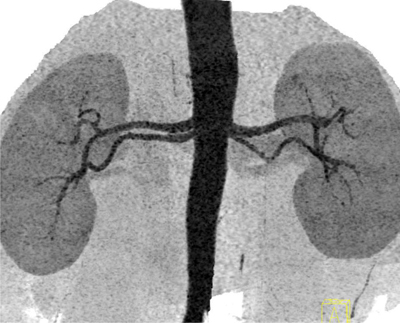
The aim of our study was to determine the efficacy of preoperative early arterial Computed tomography angiography (CTA) in donor nephrectomy, to assess the renal arterial and venous structures of donor kidneys.

MATERIALS AND METHODS

Seventy living donor candidates were included to this study, who had CTA for the assessment of their renal vessels in our hospital between January 2011 and January 2015. Only early arterial phase images were obtained to avoid exposing the patients from high dose of radiation. Scans were reported by two radiologists independently. The number of renal arteries, veins and their tributaries were documented. The donor kidneys were removed by two consultant surgeons, and after back-table perfusion the same details were recorded and taken as the reference findings for the operation side.

RESULTS

A total of 70 potential live kidney donors underwent renal CTA, among them fifty five patients had donor nephrectomy. A total of 140 kidneys were evaluated by CTA and the vessels of 55 harvested kidneys were compared with CTA findings. There were 40 kidneys that had at least one accessory or polar artery. There were 5 early branching renal arteries, two retroaortic and two circumaortic renal veins. Three kidneys had multiple renal veins. Operation findings were totally consistent with CTA findings in patients who underwent donor nephrectomy.

CONCLUSION

Arterial phase CTA is sufficient for evaluation of both arterial and venous vessels of kidneys, and precontrast, venous or late phase imaging should be preserved only for chosen circumstances to avoid high radiation exposure.

摘要

目的

本研究旨在确定术前早期动脉计算机断层扫描血管造影(CTA)在供肾切除术评估供肾肾动静脉结构中的疗效。

材料与方法

本研究纳入了70例活体供肾候选者,他们于2011年1月至2015年1月期间在我院接受CTA以评估其肾血管。仅获取早期动脉期图像以避免患者受到高剂量辐射。扫描结果由两名放射科医生独立报告。记录肾动脉、静脉及其分支的数量。由两名顾问外科医生切除供肾,在后台灌注后记录相同细节,并将其作为手术侧的参考结果。

结果

共有70例潜在活体肾供者接受了肾脏CTA检查,其中55例患者接受了供肾切除术。CTA共评估了140个肾脏,并将55个摘取肾脏的血管与CTA结果进行了比较。有40个肾脏至少有一条副动脉或极动脉。有5条早期分支肾动脉,两条主动脉后肾静脉和两条主动脉周围肾静脉。3个肾脏有多条肾静脉。接受供肾切除术患者的手术结果与CTA结果完全一致。

结论

动脉期CTA足以评估肾脏的动静脉血管,仅在特定情况下应保留非增强、静脉期或延迟期成像以避免高辐射暴露。

https://cdn.ncbi.nlm.nih.gov/pmc/blobs/acd3/6450243/d970c3342825/jbsr-103-1-1719-g6.jpg
https://cdn.ncbi.nlm.nih.gov/pmc/blobs/acd3/6450243/c540ebdef0e7/jbsr-103-1-1719-g1.jpg
https://cdn.ncbi.nlm.nih.gov/pmc/blobs/acd3/6450243/73f4994df504/jbsr-103-1-1719-g2.jpg
https://cdn.ncbi.nlm.nih.gov/pmc/blobs/acd3/6450243/512a7cc56950/jbsr-103-1-1719-g3.jpg
https://cdn.ncbi.nlm.nih.gov/pmc/blobs/acd3/6450243/e868b4229a25/jbsr-103-1-1719-g4.jpg
https://cdn.ncbi.nlm.nih.gov/pmc/blobs/acd3/6450243/1fea0ec1be3b/jbsr-103-1-1719-g5.jpg
https://cdn.ncbi.nlm.nih.gov/pmc/blobs/acd3/6450243/d970c3342825/jbsr-103-1-1719-g6.jpg

相似文献

1
Evaluation of Vascular Structures of Living Donor Kidneys by Multislice Computed Tomography Angiography before Transplant Surgery: Is Arterial Phase Sufficient for Determination of Both Arteries and Veins?移植手术前多层螺旋计算机断层血管造影对活体供肾血管结构的评估:动脉期对动静脉的判定是否足够?
J Belg Soc Radiol. 2019 Apr 4;103(1):23. doi: 10.5334/jbsr.1719.
2
A prospective study of the predictive power of spiral computed tomographic angiography for defining renal vascular anatomy before live-donor nephrectomy.一项关于螺旋计算机断层血管造影术在活体供肾肾切除术前行肾血管解剖定义预测能力的前瞻性研究。
BJU Int. 2004 Nov;94(7):1077-81. doi: 10.1111/j.1464-410X.2004.05107.x.
3
Assessment of 100 live potential renal donors for laparoscopic nephrectomy with multi-detector row helical CT.采用多排螺旋CT对100名潜在活体肾供体进行腹腔镜肾切除术评估。
Radiology. 2005 Dec;237(3):973-80. doi: 10.1148/radiol.2373041303.
4
Evaluation of the Renal Arteries of 2,144 Living Kidney Donors Using Computed Tomography Angiography and Comparison with Intraoperative Findings.应用计算机断层血管造影术评估 2144 名活体供肾者的肾动脉,并与术中所见进行比较。
Urol Int. 2020;104(7-8):637-640. doi: 10.1159/000507796. Epub 2020 May 14.
5
Prospective study comparing three-dimensional computed tomography and magnetic resonance imaging for evaluating the renal vascular anatomy in potential living renal donors.比较三维计算机断层扫描和磁共振成像在评估潜在活体肾供体肾血管解剖结构中的前瞻性研究。
BJU Int. 2005 Nov;96(7):1105-8. doi: 10.1111/j.1464-410X.2005.05809.x.
6
Should the indications for laparascopic live donor nephrectomy of the right kidney be the same as for the open procedure? Anomalous left renal vasculature is not a contraindiction to laparoscopic left donor nephrectomy.右侧肾脏腹腔镜活体供肾切除术的指征应与开放手术相同吗?异常的左肾血管并非腹腔镜左供肾切除术的禁忌证。
Transplantation. 2001 Mar 15;71(5):660-4. doi: 10.1097/00007890-200103150-00015.
7
Diagnostic accuracy of 64-MDCT angiography in the preoperative evaluation of renal vessels and compared with laparotomy findings in living donor kidney.64 层 MDCT 血管造影术在活体供肾术前评估肾血管中的诊断准确性,并与剖腹探查结果比较。
Ren Fail. 2014 Apr;36(3):327-31. doi: 10.3109/0886022X.2013.862768. Epub 2013 Dec 2.
8
Multidetector computed tomography for preoperative evaluation of vascular anatomy in living renal donors.多层螺旋计算机断层扫描在活体肾供体血管解剖术前评估中的应用
Surg Radiol Anat. 2009 Apr;31(4):227-35. doi: 10.1007/s00276-008-0428-0. Epub 2008 Oct 25.
9
Comprehensive MR imaging in the preoperative evaluation of living donor candidates for laparoscopic nephrectomy: initial experience.磁共振成像在腹腔镜肾切除术活体供肾者术前评估中的综合应用:初步经验
Radiology. 2002 Nov;225(2):427-32. doi: 10.1148/radiol.2252011671.
10
Noncontrast-Enhanced Magnetic Resonance Versus Computed Tomography Angiography in Preoperative Evaluation of Potential Living Renal Donors.非增强磁共振成像与计算机断层血管造影术在潜在活体肾供体术前评估中的比较
Acad Radiol. 2015 Nov;22(11):1368-75. doi: 10.1016/j.acra.2015.06.015. Epub 2015 Aug 22.

引用本文的文献

1
Application of low-dose CT to the creation of 3D-printed kidney and perinephric tissue models for laparoscopic nephrectomy.低剂量 CT 在腹腔镜肾切除术 3D 打印肾及肾周组织模型创建中的应用。
Cancer Med. 2021 May;10(9):3077-3084. doi: 10.1002/cam4.3851. Epub 2021 Apr 2.
2
Complex Surgical Reconstruction of Upper Pole Artery in Living-Donor Kidney Transplantation.活体供肾移植中肾上极动脉的复杂外科重建。
Ann Transplant. 2021 Jan 15;26:e926850. doi: 10.12659/AOT.926850.

本文引用的文献

1
Renal volume assessed by magnetic resonance imaging volumetry correlates with renal function in living kidney donors pre- and postdonation: a retrospective cohort study.磁共振成像容积测量评估的肾体积与活体供肾者捐赠前后的肾功能相关:一项回顾性队列研究。
Transpl Int. 2018 Jul;31(7):773-780. doi: 10.1111/tri.13150. Epub 2018 Apr 15.
2
Quantification of Single-Kidney Function and Volume in Living Kidney Donors Using Dynamic Contrast-Enhanced MRI.应用动态对比增强 MRI 对活体供肾者单肾功能和体积进行定量评估。
AJR Am J Roentgenol. 2016 Nov;207(5):1022-1030. doi: 10.2214/AJR.16.16168. Epub 2016 Aug 24.
3
3.0Tesla magnetic resonance angiography (MRA) for comprehensive renal evaluation of living renal donors: pilot study with computerized tomography angiography (CTA) comparison.
3.0特斯拉磁共振血管造影术(MRA)用于活体肾供体的肾脏全面评估:与计算机断层扫描血管造影术(CTA)比较的初步研究
Clin Imaging. 2016 May-Jun;40(3):370-7. doi: 10.1016/j.clinimag.2016.01.010. Epub 2016 Jan 23.
4
Renal blood flow using arterial spin labelling MRI and calculated filtration fraction in healthy adult kidney donors Pre-nephrectomy and post-nephrectomy.使用动脉自旋标记磁共振成像(MRI)测量健康成年肾供体术前和术后的肾血流量,并计算滤过分数。
Eur Radiol. 2015 Aug;25(8):2390-6. doi: 10.1007/s00330-015-3594-6. Epub 2015 Feb 11.
5
Role of multidetector computed tomography for evaluation of living kidney donors.多排螺旋计算机断层扫描在活体肾供体评估中的作用。
Nephrourol Mon. 2013 Sep;5(4):870-3. doi: 10.5812/numonthly.10875. Epub 2013 Aug 12.
6
Does kidney transplantation with multiple arteries affect graft survival?多支肾动脉的肾移植会影响移植物存活吗?
Transplant Proc. 2011 Nov;43(9):3423-5. doi: 10.1016/j.transproceed.2011.09.027.
7
Vascular constraints in laparoscopic renal allograft: comparative analysis of multiple and single renal arteries in 976 laparoscopic donor nephrectomies.腹腔镜肾移植中的血管限制:976 例腹腔镜供肾切术中多发性和单发性肾动脉的对比分析。
World J Surg. 2011 Sep;35(9):2159-66. doi: 10.1007/s00268-011-1168-6.
8
Multidetector CT of living renal donors: lessons learned from surgeons.多排螺旋 CT 活体供肾者:外科医生的经验教训。
Radiographics. 2010 Nov;30(7):1875-90. doi: 10.1148/rg.307105032.
9
The utility of 64 channel multidetector CT angiography for evaluating the renal vascular anatomy and possible variations: a pictorial essay.64 通道多排螺旋 CT 血管造影在评估肾血管解剖及可能变异中的应用:影像学研究。
Korean J Radiol. 2010 May-Jun;11(3):346-54. doi: 10.3348/kjr.2010.11.3.346. Epub 2010 Apr 29.
10
CT angiography of the renal arteries and veins: normal anatomy and variants.肾动脉和静脉 CT 血管造影:正常解剖结构和变异。
Diagn Interv Radiol. 2011 Mar;17(1):67-73. doi: 10.4261/1305-3825.DIR.2902-09.1. Epub 2010 Feb 9.