• 文献检索
  • 文档翻译
  • 深度研究
  • 学术资讯
  • Suppr Zotero 插件Zotero 插件
  • 邀请有礼
  • 套餐&价格
  • 历史记录
应用&插件
Suppr Zotero 插件Zotero 插件浏览器插件Mac 客户端Windows 客户端微信小程序
定价
高级版会员购买积分包购买API积分包
服务
文献检索文档翻译深度研究API 文档MCP 服务
关于我们
关于 Suppr公司介绍联系我们用户协议隐私条款
关注我们

Suppr 超能文献

核心技术专利:CN118964589B侵权必究
粤ICP备2023148730 号-1Suppr @ 2026

文献检索

告别复杂PubMed语法,用中文像聊天一样搜索,搜遍4000万医学文献。AI智能推荐,让科研检索更轻松。

立即免费搜索

文件翻译

保留排版,准确专业,支持PDF/Word/PPT等文件格式,支持 12+语言互译。

免费翻译文档

深度研究

AI帮你快速写综述,25分钟生成高质量综述,智能提取关键信息,辅助科研写作。

立即免费体验

64 通道多排螺旋 CT 血管造影在评估肾血管解剖及可能变异中的应用:影像学研究。

The utility of 64 channel multidetector CT angiography for evaluating the renal vascular anatomy and possible variations: a pictorial essay.

机构信息

Department of Radiodiagnosis, Sanjay Gandhi Post Graduate Institute of Medical Sciences, Lucknow, Uttar Pradesh, India.

出版信息

Korean J Radiol. 2010 May-Jun;11(3):346-54. doi: 10.3348/kjr.2010.11.3.346. Epub 2010 Apr 29.

DOI:10.3348/kjr.2010.11.3.346
PMID:20461189
原文链接:https://pmc.ncbi.nlm.nih.gov/articles/PMC2864862/
Abstract

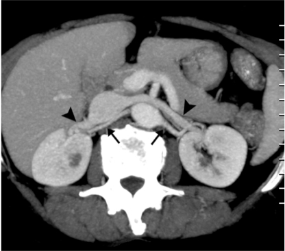
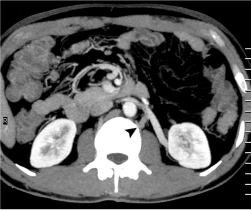
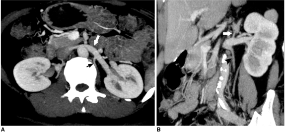
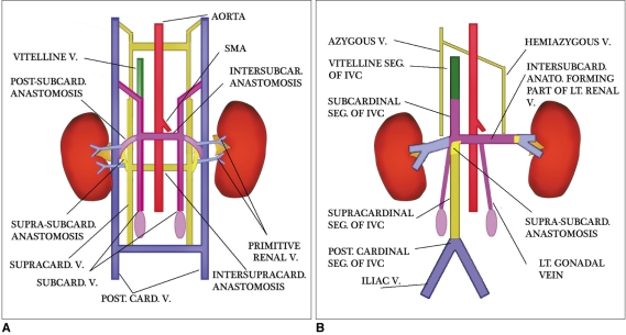
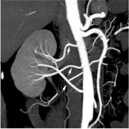
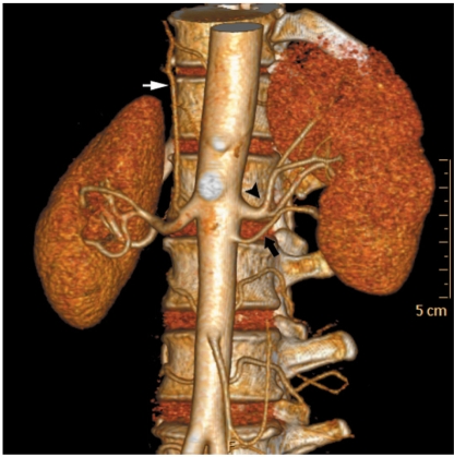
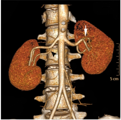
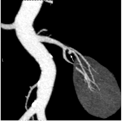
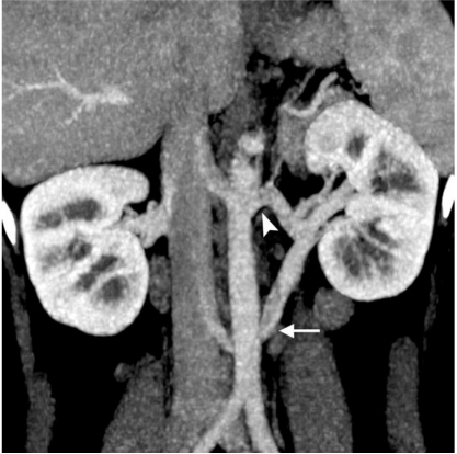
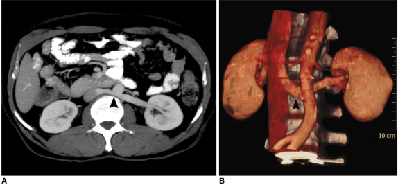
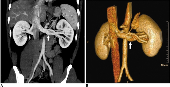
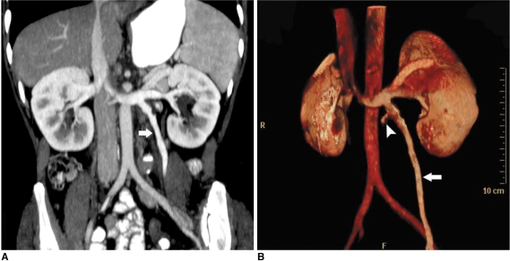
The increased use of laparoscopic nephrectomy and nephron-sparing surgery has prompted the need for a more detailed radiological evaluation of the renal vascular anatomy. Multidetector CT angiography is a fast and accurate modality for assessing the precise anatomy of the renal vessels. In this pictorial review, we present the multidetector CT angiography appearances of the normal renal vascular anatomy and a spectrum of various anomalies that require accurate vascular depiction before undergoing surgical treatment.

摘要

腹腔镜肾切除术和保肾手术的应用日益增多,这促使我们需要更详细地评估肾脏血管解剖结构。多层螺旋 CT 血管造影术是一种快速准确的评估肾脏血管精确解剖结构的方法。在本影像学综述中,我们展示了多层螺旋 CT 血管造影术对正常肾脏血管解剖结构的表现以及一系列需要在手术治疗前准确描述血管的各种异常。

https://cdn.ncbi.nlm.nih.gov/pmc/blobs/6f55/2864862/09e7f0970662/kjr-11-346-g017.jpg
https://cdn.ncbi.nlm.nih.gov/pmc/blobs/6f55/2864862/8fe7a5cdbca2/kjr-11-346-g001.jpg
https://cdn.ncbi.nlm.nih.gov/pmc/blobs/6f55/2864862/3c7e9b1fd5c7/kjr-11-346-g002.jpg
https://cdn.ncbi.nlm.nih.gov/pmc/blobs/6f55/2864862/53667fac874b/kjr-11-346-g003.jpg
https://cdn.ncbi.nlm.nih.gov/pmc/blobs/6f55/2864862/d32ec238f23f/kjr-11-346-g004.jpg
https://cdn.ncbi.nlm.nih.gov/pmc/blobs/6f55/2864862/5c3269b2f8bd/kjr-11-346-g005.jpg
https://cdn.ncbi.nlm.nih.gov/pmc/blobs/6f55/2864862/dc78f06b0967/kjr-11-346-g006.jpg
https://cdn.ncbi.nlm.nih.gov/pmc/blobs/6f55/2864862/8e299d4a24e0/kjr-11-346-g007.jpg
https://cdn.ncbi.nlm.nih.gov/pmc/blobs/6f55/2864862/20936474b76b/kjr-11-346-g008.jpg
https://cdn.ncbi.nlm.nih.gov/pmc/blobs/6f55/2864862/d9b0f0097fc0/kjr-11-346-g009.jpg
https://cdn.ncbi.nlm.nih.gov/pmc/blobs/6f55/2864862/a341ce92fd4b/kjr-11-346-g010.jpg
https://cdn.ncbi.nlm.nih.gov/pmc/blobs/6f55/2864862/07c164b88359/kjr-11-346-g011.jpg
https://cdn.ncbi.nlm.nih.gov/pmc/blobs/6f55/2864862/8d442220a5f0/kjr-11-346-g012.jpg
https://cdn.ncbi.nlm.nih.gov/pmc/blobs/6f55/2864862/e8bfcd9710fe/kjr-11-346-g013.jpg
https://cdn.ncbi.nlm.nih.gov/pmc/blobs/6f55/2864862/6768bfca43c3/kjr-11-346-g014.jpg
https://cdn.ncbi.nlm.nih.gov/pmc/blobs/6f55/2864862/1e217abb3477/kjr-11-346-g015.jpg
https://cdn.ncbi.nlm.nih.gov/pmc/blobs/6f55/2864862/8ebb33ce0def/kjr-11-346-g016.jpg
https://cdn.ncbi.nlm.nih.gov/pmc/blobs/6f55/2864862/09e7f0970662/kjr-11-346-g017.jpg

相似文献

1
The utility of 64 channel multidetector CT angiography for evaluating the renal vascular anatomy and possible variations: a pictorial essay.64 通道多排螺旋 CT 血管造影在评估肾血管解剖及可能变异中的应用:影像学研究。
Korean J Radiol. 2010 May-Jun;11(3):346-54. doi: 10.3348/kjr.2010.11.3.346. Epub 2010 Apr 29.
2
Multidetector CT angiography of renal vasculature: normal anatomy and variants.肾脏血管系统的多排螺旋CT血管造影:正常解剖结构及变异
Eur Radiol. 2009 Jan;19(1):236-44. doi: 10.1007/s00330-008-1126-3. Epub 2008 Jul 30.
3
CT angiography of the renal arteries and veins: normal anatomy and variants.肾动脉和静脉 CT 血管造影:正常解剖结构和变异。
Diagn Interv Radiol. 2011 Mar;17(1):67-73. doi: 10.4261/1305-3825.DIR.2902-09.1. Epub 2010 Feb 9.
4
Multidetector CT angiography for preoperative evaluation of living laparoscopic kidney donors.多排螺旋CT血管造影术用于活体腹腔镜肾供体的术前评估。
AJR Am J Roentgenol. 2003 Jun;180(6):1633-8. doi: 10.2214/ajr.180.6.1801633.
5
Helical computerized tomography arteriography for evaluation of live renal donors undergoing laparoscopic nephrectomy.螺旋计算机断层扫描血管造影术用于评估接受腹腔镜肾切除术的活体肾供体。
J Urol. 1999 Jul;162(1):31-4. doi: 10.1097/00005392-199907000-00008.
6
Diagnostic accuracy of a volume-rendered computed tomography movie and other computed tomography-based imaging methods in assessment of renal vascular anatomy for laparoscopic donor nephrectomy.容积再现 CT 电影与其他 CT 成像方法在评估腹腔镜供肾切取术肾血管解剖中的诊断准确性。
Int Urol Nephrol. 2009 Dec;41(4):785-90. doi: 10.1007/s11255-009-9535-z. Epub 2009 Mar 6.
7
Multidetector computed tomography for preoperative evaluation of vascular anatomy in living renal donors.多层螺旋计算机断层扫描在活体肾供体血管解剖术前评估中的应用
Surg Radiol Anat. 2009 Apr;31(4):227-35. doi: 10.1007/s00276-008-0428-0. Epub 2008 Oct 25.
8
Multidetector CT angiography in living donor renal transplantation: accuracy and discrepancies in right venous anatomy.多排 CT 血管造影在活体供肾移植中的应用:右静脉解剖的准确性和差异。
Clin Transplant. 2011 Jan-Feb;25(1):77-82. doi: 10.1111/j.1399-0012.2009.01193.x.
9
Role of CT angiography in the preoperative evaluation for laparoscopic nephrectomy.CT血管造影在腹腔镜肾切除术术前评估中的作用。
Radiographics. 1998 May-Jun;18(3):589-601. doi: 10.1148/radiographics.18.3.9599384.
10
3-dimensional volume rendered computerized tomography for preoperative evaluation and intraoperative treatment of patients undergoing nephron sparing surgery.三维容积再现计算机断层扫描在保留肾单位手术患者术前评估及术中治疗中的应用
J Urol. 1999 Apr;161(4):1097-102.

引用本文的文献

1
The accuracy of a new noninvasive approach using physical examination combined with ultrasonography to detect intraabdominal hypertension.一种采用体格检查联合超声检查来检测腹腔内高压的新型非侵入性方法的准确性。
Sci Rep. 2025 Jul 15;15(1):25488. doi: 10.1038/s41598-025-10888-0.
2
Enhancing procedural decision making with cone beam CT in renal artery embolization.在肾动脉栓塞中使用锥形束 CT 增强手术决策。
Sci Rep. 2024 Aug 6;14(1):18198. doi: 10.1038/s41598-024-69363-x.
3
Protean Drainage Patterns of the Left Renal Vein: A Cadaveric and Retrospective Clinical Study on the Surgical Implications and Technical Feasibility.

本文引用的文献

1
Double inferior vena cava: a report of three cases.双下腔静脉:三例报告
Singapore Med J. 2009 Jun;50(6):e211-3.
2
CT angiography for living kidney donors: accuracy, cause of misinterpretation and prevalence of variation.活体肾供体的CT血管造影:准确性、误判原因及变异发生率
Korean J Radiol. 2008 Jul-Aug;9(4):333-9. doi: 10.3348/kjr.2008.9.4.333.
3
Imaging the inferior vena cava: a road less traveled.下腔静脉成像:一条少有人走的路。
左肾静脉的多种引流模式:关于手术意义和技术可行性的尸体解剖及回顾性临床研究
Cureus. 2024 Jun 24;16(6):e63037. doi: 10.7759/cureus.63037. eCollection 2024 Jun.
4
Photon-counting CT: technical features and clinical impact on abdominal imaging.光子计数 CT:腹部成像的技术特点和临床影响。
Abdom Radiol (NY). 2024 Dec;49(12):4383-4399. doi: 10.1007/s00261-024-04414-5. Epub 2024 Jun 18.
5
Vascular Variations and Incidental Pathologies in Potential Living Renal Donors Using 160-Slice Multidetector Computed Tomography Angiography.使用160层多层螺旋计算机断层血管造影术对潜在活体肾供体的血管变异和偶然病变进行研究。
Cureus. 2023 Jul 7;15(7):e41502. doi: 10.7759/cureus.41502. eCollection 2023 Jul.
6
Evaluation of Vascular Structures of Living Donor Kidneys by Multislice Computed Tomography Angiography before Transplant Surgery: Is Arterial Phase Sufficient for Determination of Both Arteries and Veins?移植手术前多层螺旋计算机断层血管造影对活体供肾血管结构的评估:动脉期对动静脉的判定是否足够?
J Belg Soc Radiol. 2019 Apr 4;103(1):23. doi: 10.5334/jbsr.1719.
7
Development of the renal vasculature.肾脏血管系统的发育。
Semin Cell Dev Biol. 2019 Jul;91:132-146. doi: 10.1016/j.semcdb.2018.06.001. Epub 2018 Jun 6.
8
Dual Retro-Aortic Left Renal Vein with Drainage into Left Common Iliac Vein: Rare Anomaly of Left Renal Vein.双后主动脉左肾静脉并引流至左髂总静脉:左肾静脉的罕见异常
Int J Organ Transplant Med. 2016;7(2):99-101. Epub 2016 May 1.
9
A rare anomaly of left renal vein drainage into the left common iliac vein: A case report.左肾静脉引流至左髂总静脉的罕见异常:一例报告。
Int J Surg Case Rep. 2016;20:4-6. doi: 10.1016/j.ijscr.2015.12.050. Epub 2016 Jan 7.
10
Are spinal or paraspinal anatomic markers helpful for vertebral numbering and diagnosing lumbosacral transitional vertebrae?脊柱或脊柱旁解剖标志物对腰椎骶化的椎体编号和诊断有帮助吗?
Korean J Radiol. 2014 Mar-Apr;15(2):258-66. doi: 10.3348/kjr.2014.15.2.258. Epub 2014 Mar 7.
Radiographics. 2008 May-Jun;28(3):669-89. doi: 10.1148/rg.283075101.
4
Surgically relevant normal and variant renal parenchymal and vascular anatomy in preoperative 16-MDCT evaluation of potential laparoscopic renal donors.潜在腹腔镜肾供体术前16层MDCT评估中与手术相关的正常及变异肾实质和血管解剖结构
AJR Am J Roentgenol. 2007 Jan;188(1):105-14. doi: 10.2214/AJR.05.1002.
5
Renal artery origins and variations: angiographic evaluation of 855 consecutive patients.肾动脉起源及变异:855例连续患者的血管造影评估
Diagn Interv Radiol. 2006 Dec;12(4):183-6.
6
Utility of 16-MDCT angiography for comprehensive preoperative vascular evaluation of laparoscopic renal donors.16层螺旋CT血管造影术在腹腔镜肾供体术前全面血管评估中的应用价值
AJR Am J Roentgenol. 2006 Jun;186(6):1630-8. doi: 10.2214/AJR.05.0983.
7
Assessment of 100 live potential renal donors for laparoscopic nephrectomy with multi-detector row helical CT.采用多排螺旋CT对100名潜在活体肾供体进行腹腔镜肾切除术评估。
Radiology. 2005 Dec;237(3):973-80. doi: 10.1148/radiol.2373041303.
8
Evaluation of the renal venous system on late arterial and venous phase images with MDCT angiography in potential living laparoscopic renal donors.在潜在的活体腹腔镜肾供体中,利用多层螺旋CT血管造影术在动脉晚期和静脉期图像上评估肾静脉系统。
AJR Am J Roentgenol. 2005 Feb;184(2):539-45. doi: 10.2214/ajr.184.2.01840539.
9
Techniques, clinical applications and limitations of 3D reconstruction in CT of the abdomen.腹部CT中三维重建的技术、临床应用及局限性
Korean J Radiol. 2004 Jan-Mar;5(1):55-67. doi: 10.3348/kjr.2004.5.1.55.
10
Three-dimensional volume-rendered CT angiography of the renal arteries and veins: normal anatomy, variants, and clinical applications.肾动脉和肾静脉的三维容积再现CT血管造影:正常解剖结构、变异及临床应用
Radiographics. 2001 Mar-Apr;21(2):373-86; questionnaire 549-55. doi: 10.1148/radiographics.21.2.g01mr19373.