Pei Jing, Zhang Jing, Yang Xiaowei, Wu Zhengsheng, Sun Chenyun, Wang Zhaorui, Wang Benzhong
1Department of Breast Surgery, Department of General Surgery, The First Affiliated Hospital of Anhui Medical University, Number 218, Jixi Road, Hefei, 230022 Anhui People's Republic of China.
Department of Breast Surgery, The Tumor Hospital of XuZhou, HuanCheng Road 131, Xuzhou, 221003 Jiangsu People's Republic of China.
Cancer Cell Int. 2019 Mar 29;19:75. doi: 10.1186/s12935-019-0791-4. eCollection 2019.
The role of TMED3 involved in cancers has been seldom described, let alone in breast cancer. To explore the clinicopathological significance of TMED3 expression and the biological roles involved in breast cancer cells, we undertook the study.
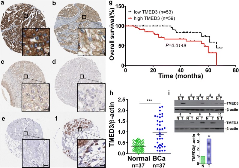
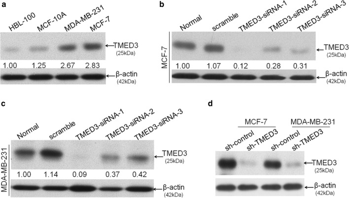
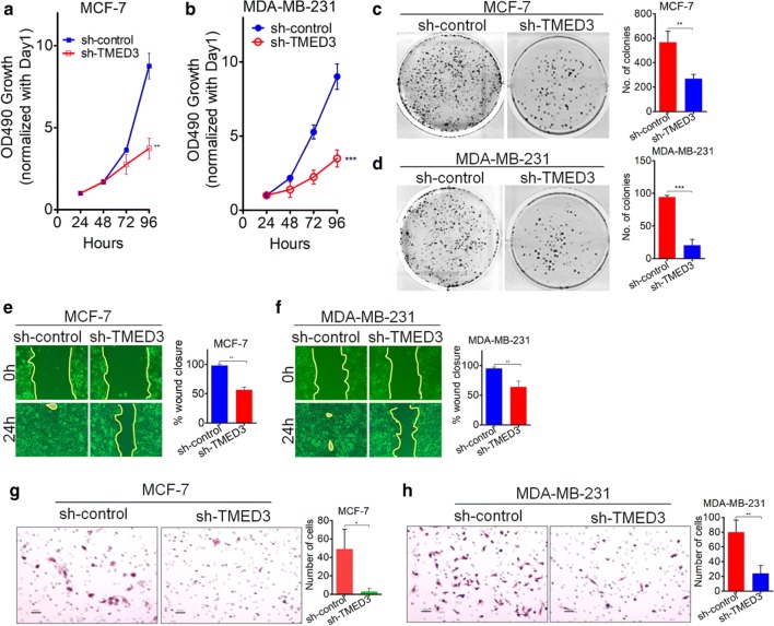
Immunohistochemistry was performed to observe the pattern of TMED3 expression in breast cancer tissues, totaling 224 cases; followed by detailed statistical analysis between TMED3 expression versus clinicopathological information available. To explore the role of TMED3 involved in the malignant behaviors of breast cancer cells, wound-healing and Transwell assays were conducted to evaluate the variation of migration and invasion of MCF-7 and MDA-MB-231 cells whose TMED3 has been stably silenced using lenti-viral based short hairpin RNA (shRNA) vectors. MTT, clonogenic assay and xenograft nude mice model were undertaken to observe the variation of proliferation both in vitro and in vivo.
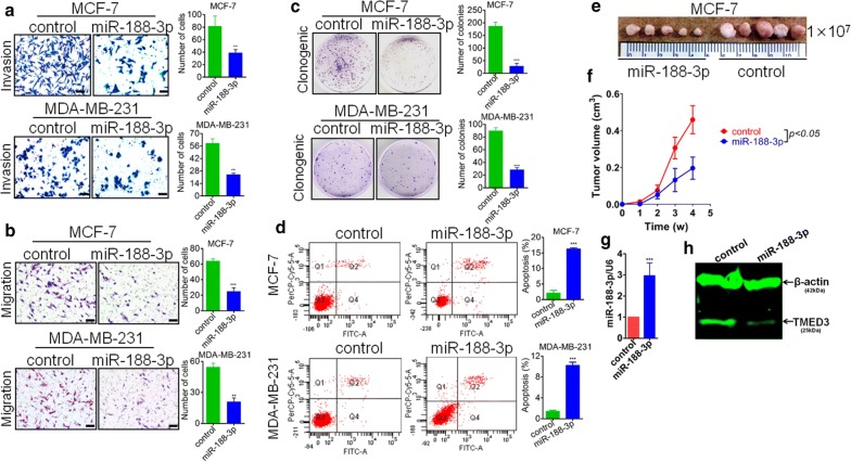
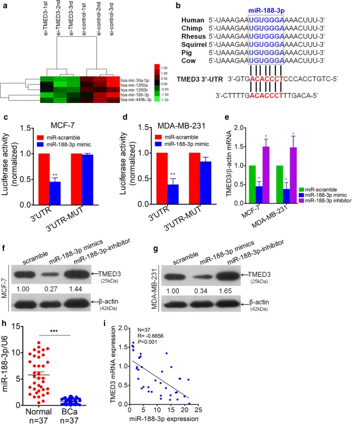
It was shown that elevated TMED3 markedly correlated with ER, PR, Her-2 status, and lymph nodes metastases in addition to significant association with poor overall prognosis. In vitro, TMED3 was shown to promote proliferation, migration and invasion of breast cancer cells. Moreover, miR-188-3p was identified as a novel negative regulator of TMED3 in breast cancer, which can slow down the proliferation, migration and invasion of MCF-7 cells. Results from in vivo xenograft nude mice models showed that lenti-viral based miR-188-3p re-expression can markedly impair the tumor growth.
Our data define and bolster the oncogenic role of TMED3 in breast cancer.
TMED3在癌症中的作用鲜有报道,在乳腺癌中更是如此。为了探究TMED3表达的临床病理意义及其在乳腺癌细胞中的生物学作用,我们开展了本研究。
对224例乳腺癌组织进行免疫组化,观察TMED3的表达模式;随后对TMED3表达与现有临床病理信息进行详细的统计分析。为了探究TMED3在乳腺癌细胞恶性行为中的作用,采用基于慢病毒的短发夹RNA(shRNA)载体稳定沉默TMED3,通过伤口愈合实验和Transwell实验评估MCF-7和MDA-MB-231细胞迁移和侵袭能力的变化。采用MTT实验、克隆形成实验和异种移植裸鼠模型观察体外和体内增殖能力的变化。
结果显示,TMED3表达升高不仅与ER、PR、Her-2状态及淋巴结转移显著相关,还与总体预后不良密切相关。在体外,TMED3可促进乳腺癌细胞的增殖、迁移和侵袭。此外,miR-188-3p被鉴定为乳腺癌中TMED3的新型负调控因子,可减缓MCF-7细胞的增殖、迁移和侵袭。体内异种移植裸鼠模型结果显示,基于慢病毒的miR-188-3p重新表达可显著抑制肿瘤生长。
我们的数据明确并支持了TMED3在乳腺癌中的致癌作用。