Lee You Jeong, Ahn Eun-Young, Park Youmie
College of Pharmacy and Inje Institute of Pharmaceutical Sciences and Research, Inje University, 197 Inje-ro, Gimhae, Gyeongnam, 50834, Republic of Korea.
Nanoscale Res Lett. 2019 Apr 11;14(1):129. doi: 10.1186/s11671-019-2967-1.
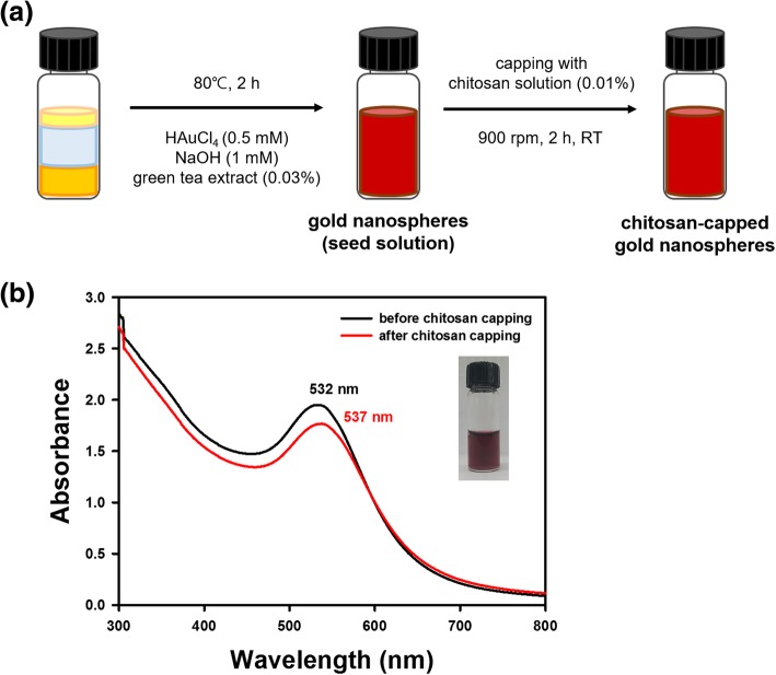
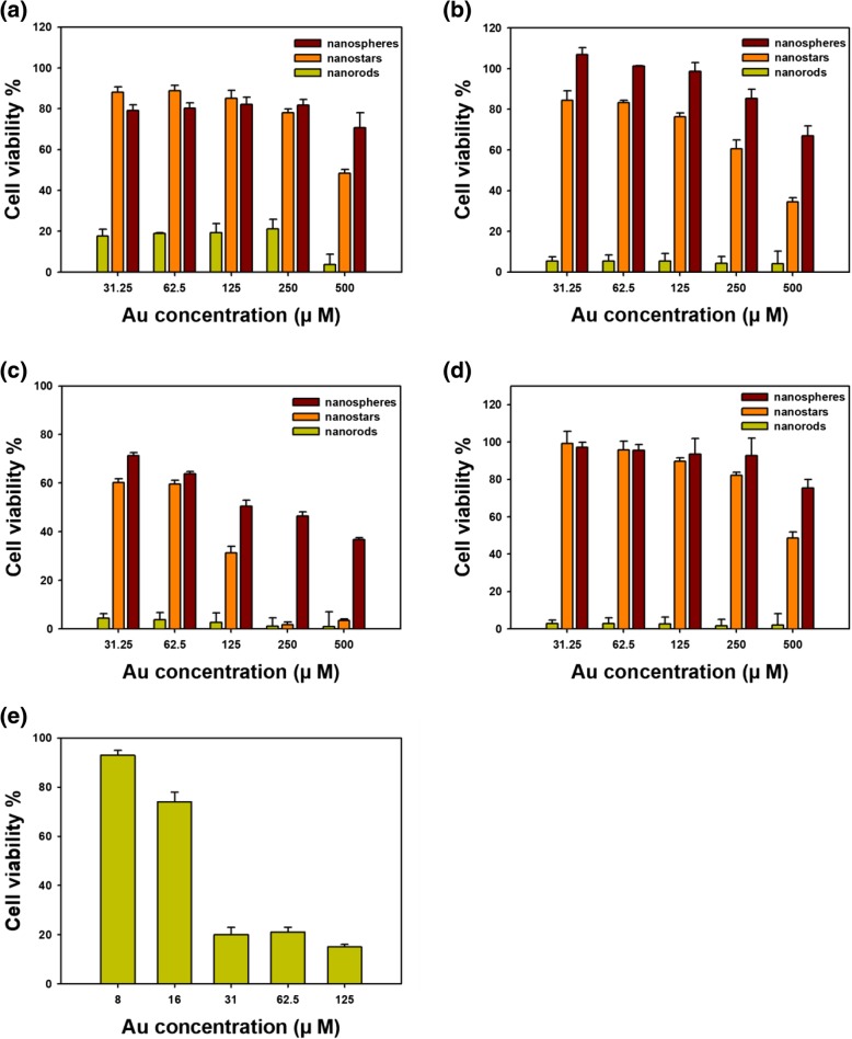
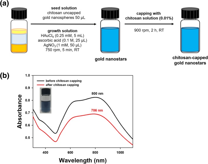
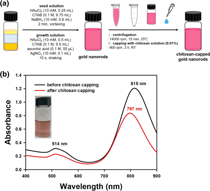
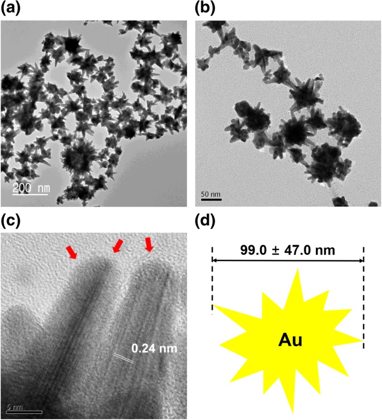
In the present report, three different shapes of chitosan-capped gold nanoparticles (nanospheres, nanostars, and nanorods) were synthesized to investigate the effects of shape on cytotoxicity and cellular uptake in cancer cells. Green tea extract was utilized as a reducing agent to reduce gold salts to gold nanospheres. Gold nanostars were prepared using an as-prepared nanosphere solution as a seed solution. Gold nanorods were synthesized using a conventional method. All three types of gold nanoparticles showed their characteristic surface plasmon resonance bands upon UV-visible spectrophotometry. In high-resolution transmission electron microscopy images, lattice structures were clearly observed in all three shapes, confirming the crystalline nature of the nanoparticles. All three colloidal solutions of gold nanoparticles retained colloidal stability in various solutions. To assess cytotoxicity, the 3-(4,5-dimethylthiazol-2-yl)-2,5-diphenyltetrazolium bromide (MTT) assay was performed on four cancer cell lines. The cytotoxicity was the highest in nanorods, followed by nanostars and finally nanospheres. The cellular uptake of gold nanoparticles in human hepatocyte carcinoma cells (HepG2) was measured, and the results followed the order nanospheres > nanorods > nanostars. The outcomes of the current study may assist in the shape design of gold nanoparticles for therapeutic applications as drug delivery vehicles in the field of nanomedicine.
在本报告中,合成了三种不同形状的壳聚糖包覆金纳米颗粒(纳米球、纳米星和纳米棒),以研究形状对癌细胞的细胞毒性和细胞摄取的影响。利用绿茶提取物作为还原剂将金盐还原为金纳米球。使用制备好的纳米球溶液作为种子溶液制备金纳米星。采用常规方法合成金纳米棒。在紫外-可见分光光度法下,所有三种类型的金纳米颗粒均显示出其特征性的表面等离子体共振带。在高分辨率透射电子显微镜图像中,在所有三种形状中均清晰观察到晶格结构,证实了纳米颗粒的晶体性质。金纳米颗粒的所有三种胶体溶液在各种溶液中均保持胶体稳定性。为了评估细胞毒性,对四种癌细胞系进行了3-(4,5-二甲基噻唑-2-基)-2,5-二苯基四氮唑溴盐(MTT)测定。细胞毒性在纳米棒中最高,其次是纳米星,最后是纳米球。测定了金纳米颗粒在人肝癌细胞(HepG2)中的细胞摄取情况,结果顺序为纳米球>纳米棒>纳米星。本研究结果可能有助于在纳米医学领域设计用于治疗应用的金纳米颗粒形状,作为药物递送载体。