Ho Cheng-Maw, Ho Shu-Li, Jeng Yung-Ming, Lai Yu-Sheng, Chen Ya-Hui, Lu Shao-Chun, Chen Hui-Ling, Chang Po-Yuan, Hu Rey-Heng, Lee Po-Huang
1Department of Surgery, National Taiwan University Hospital and College of Medicine, Taipei, Taiwan.
2Hepatitis Research Center, National Taiwan University Hospital, 7 Chung-Shan South Road, Taipei, 100 Taiwan.
J Inflamm (Lond). 2019 Apr 2;16:7. doi: 10.1186/s12950-019-0211-5. eCollection 2019.
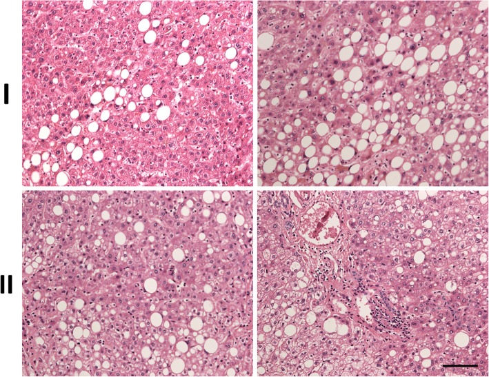
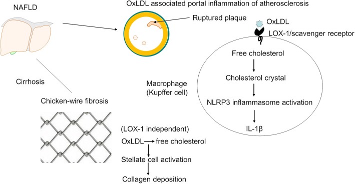
Macrophages engulf oxidized-LDL (oxLDL) leading to accumulation of cellular cholesterol and formation of foam cells, which is a hallmark of atherosclerosis. Moreover, recent studies showed that accumulation of free cholesterol in macrophages leading to activation of NLRP3 inflammasome and production of interleukin-1β (IL-1β) has been linked to atherosclerosis-associated inflammation. However, it is not clear if cholesterol accumulation is associated with hepatic inflammation and fibrosis in the liver. In this study, we investigated the association of free cholesterol and oxLDL accumulation in portal vein with the inflammation, atherosclerosis, and fibrosis in human nonalcoholic fatty liver disease (NAFLD).
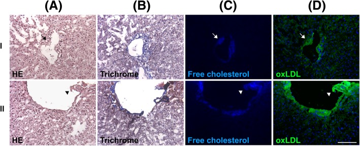
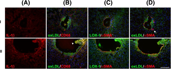
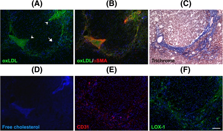
Serial sections derived from surgical specimens of NAFLD were stained with filipin and antibodies against IL-1β, CD68, α-smooth muscle actin (α-SMA), oxLDL and lectin-like oxLDL receptor-1 (LOX-1).
We show that free cholesterol was colocalized with oxLDL in the wall of portal vein, and which was associated with lumen narrowing, plaque formation, endothelium deformation, and portal venous inflammation. The inflammation was evidenced by the colocalization of Kupffer cells and IL-1β and the expression of LOX-1. Notably, ruptured plaque was closely associated with portal venous inflammation. Moreover, free cholesterol and oxLDL accumulation in periportal and sinusoidal fibrosis, which was associated with regional stellate cell activation and chicken-wire fibrosis.
These findings reveal a direct association between cholesterol accumulation, portal venous inflammation and fibrosis in NAFLD.
巨噬细胞吞噬氧化低密度脂蛋白(oxLDL)会导致细胞胆固醇积聚并形成泡沫细胞,这是动脉粥样硬化的一个标志。此外,最近的研究表明,巨噬细胞中游离胆固醇的积聚导致NLRP3炎性小体激活和白细胞介素-1β(IL-1β)的产生,这与动脉粥样硬化相关的炎症有关。然而,尚不清楚胆固醇积聚是否与肝脏炎症和肝纤维化有关。在本研究中,我们调查了门静脉中游离胆固醇和oxLDL积聚与人类非酒精性脂肪性肝病(NAFLD)中的炎症、动脉粥样硬化和纤维化之间的关联。
对NAFLD手术标本的连续切片用制霉菌素以及抗IL-1β、CD68、α平滑肌肌动蛋白(α-SMA)、oxLDL和凝集素样oxLDL受体-1(LOX-1)的抗体进行染色。
我们发现游离胆固醇与门静脉壁中的oxLDL共定位,这与管腔狭窄、斑块形成、内皮变形和门静脉炎症有关。库普弗细胞与IL-1β的共定位以及LOX-1的表达证明了炎症的存在。值得注意的是,破裂的斑块与门静脉炎症密切相关。此外,门静脉周围和肝血窦纤维化中存在游离胆固醇和oxLDL积聚,这与局部星状细胞活化和鸡笼样纤维化有关。
这些发现揭示了NAFLD中胆固醇积聚、门静脉炎症和纤维化之间的直接关联。