Zhao Weina, Wang Xuetong, Yin Changhao, He Mengfei, Li Shuyu, Han Ying
Department of Neurology, Xuanwu Hospital of Capital Medical University, Beijing, China.
Department of Neurology, Mudanjiang Medical University Affiliated Hongqi Hospital, Mudanjiang, China.
Front Neuroinform. 2019 Mar 22;13:13. doi: 10.3389/fninf.2019.00013. eCollection 2019.
The hippocampus and hippocampal subfields have been found to be diversely affected in Alzheimer's Disease (AD) and early stages of Alzheimer's disease by neuroimaging studies. However, our knowledge is still lacking about the trajectories of the hippocampus and hippocampal subfields atrophy with the progression of Alzheimer's disease.
To identify which subfields of the hippocampus differ in the trajectories of Alzheimer's disease by magnetic resonance imaging (MRI) and to determine whether individual differences on memory could be explained by structural volumes of hippocampal subfields.
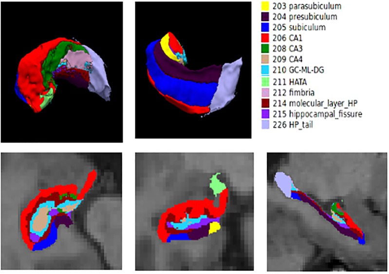
Four groups of participants including 41 AD patients, 43 amnestic mild cognitive impairment (aMCI) patients, 35 subjective cognitive decline (SCD) patients and 42 normal controls (NC) received their structural MRI brain scans. Structural MR images were processed by the FreeSurfer 6.0 image analysis suite to extract the hippocampus and its subfields. Furthermore, we investigated relationships between hippocampal subfield volumes and memory test variables (AVLT-immediate recall, AVLT-delayed recall, AVLT-recognition) and the regression model analyses were controlled for age, gender, education and eTIV.
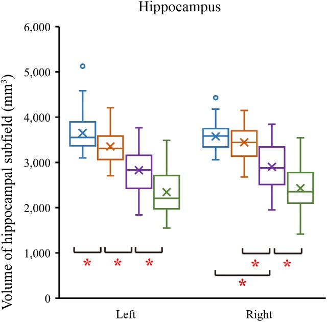
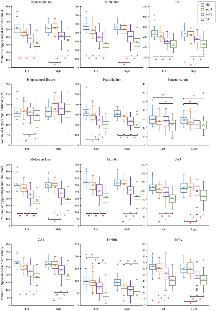
CA1, subiculum, presubiculum, molecular layer and fimbria showed the trend toward significant volume reduction among four groups with the progression of Alzheimer's disease. Volume of left subiculum was most strongly and actively correlated with performance across AVLT measures.
The trend changes in the hippocampus subfields and further illustrates that SCD is the preclinical stage of AD earlier than aMCI. Future studies should aim to associate the atrophy of the hippocampal subfields in SCD with possible conversion to aMCI or AD with longitudinal design.
神经影像学研究发现,海马体及其亚区在阿尔茨海默病(AD)和阿尔茨海默病早期受到不同程度的影响。然而,随着阿尔茨海默病的进展,我们对海马体及其亚区萎缩的轨迹仍缺乏了解。
通过磁共振成像(MRI)确定海马体的哪些亚区在阿尔茨海默病的轨迹上存在差异,并确定记忆方面的个体差异是否可以由海马体亚区的结构体积来解释。
四组参与者,包括41名AD患者、43名遗忘型轻度认知障碍(aMCI)患者、35名主观认知下降(SCD)患者和42名正常对照(NC)接受了脑部结构MRI扫描。结构MR图像由FreeSurfer 6.0图像分析套件处理,以提取海马体及其亚区。此外,我们研究了海马体亚区体积与记忆测试变量(AVLT即时回忆、AVLT延迟回忆、AVLT识别)之间的关系,并在回归模型分析中对年龄、性别、教育程度和eTIV进行了控制。
随着阿尔茨海默病的进展,CA1、下托、前下托、分子层和伞在四组中均呈现出显著体积减少的趋势。左侧下托的体积与AVLT各项测量指标的表现相关性最强且最显著。
海马体亚区的趋势变化进一步说明SCD是比aMCI更早的AD临床前期。未来的研究应旨在通过纵向设计,将SCD中海马体亚区的萎缩与可能转化为aMCI或AD联系起来。