Department of Urology, West China Hospital, Sichuan University, No. 37 Guoxue Xiang, Chengdu, 610041, Sichuan, China.
Institute of Urology, West China Hospital, Sichuan University, Chengdu, People's Republic of China, 610041.
BMC Cancer. 2019 Jan 10;19(1):49. doi: 10.1186/s12885-019-5273-5.
Due to the significant heterogeneity of renal cell carcinoma (RCC), immune checkpoints may express differently between primary and metastatic tumor. We aimed to evaluate the differential expression of TIM-3 between the primary and metastatic sites of RCC.
Cases of RCC with metastases resected or biopsied at West China Hospital between January 2009 and November 2016 were included. Clinicopathological parameters were retrospectively extracted. SPPS 22.0, GraphPad Prism 6 and R statistical software were applied for data analysis.
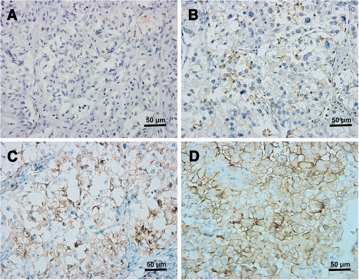
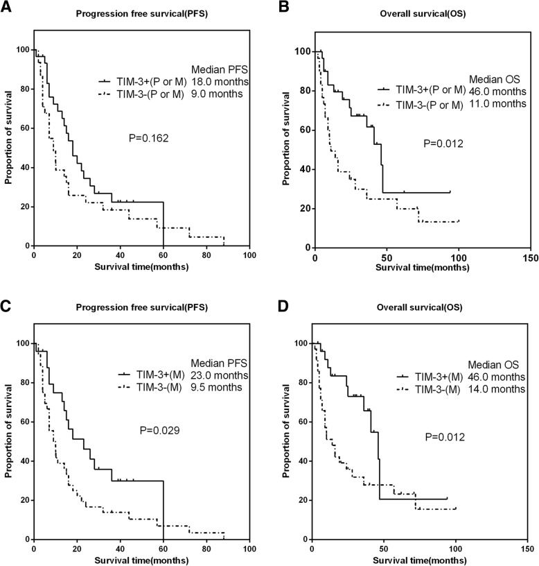
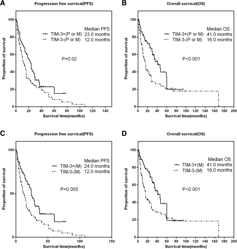
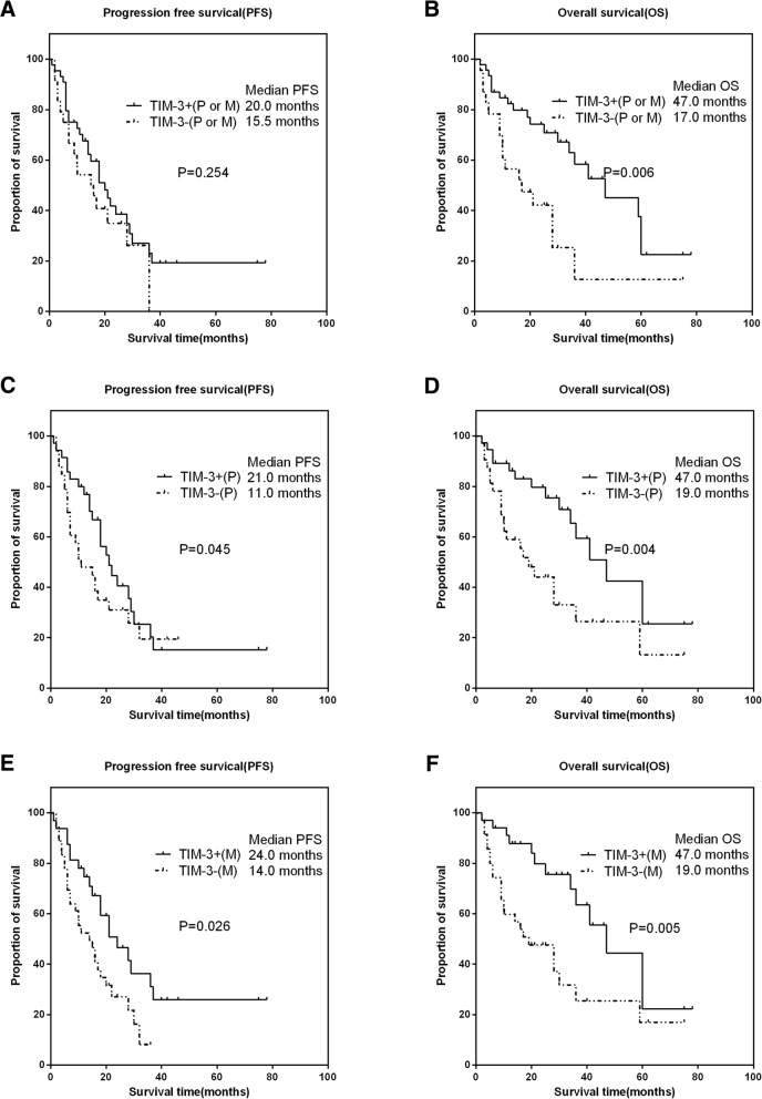
A total of 163 cases were included. Immunohistochemical results showed that the overall detection rate of TIM-3 was 56.4% (92/163). The detection rate of TIM-3 in the primary (53.0%, 44/83) was numerically higher than that of the metastasis (42.6%,79/174). Although the concordance rate of TIM-3 between the primary and metastasis was as high as 66.3% (55/83) in the paired cohort, a significant statistically difference of TIM-3 expression between the primary and metastasis was observed (χ2 = 4.664, p = 0.002), with a poor consistency (Kappa = 0.331, p = 0.002). Subsequent survival analysis suggested that TIM-3 expression either in the primary or metastatic tumor was associated with longer progression-free survival (PFS) (HR: 0.67, 95% CI 0.45-0.99, P = 0.02) and overall survival (OS) (HR: 0.52, 95% CI 0.33-0.82, P < 0.001). The expressions of TIM-3 in the primary, metastatic tumors and patients treated with targeted agents all played as favorable factors for PFS and OS. Further multivariate analysis showed that, in the whole cohort, TIM-3 expression in metastatic tumor increased the predicted accuracy (PA) of the whole model of PFS from 74.7 to 75.6% (P = 0.02). For OS, the PA of whole model was increased from 78.1 to 81.1% by adding TIM-3 expression in the metastasis (P = 0.005). The same trends were also observed in paired patients and patients treated with targeted agents. In conclusion, the expression difference between the primary and metastatic tumor of TIM-3 was significant. Biopsy or resection of the metastases may provide a more accurate biological information for clinician's decision-making and the patient's prognosis. What's more, the role of TIM-3 in the RCC still remains controversy, further study are needed to verify the conclusion.
由于肾细胞癌(RCC)的异质性显著,免疫检查点在原发肿瘤和转移肿瘤之间的表达可能不同。我们旨在评估 TIM-3 在 RCC 的原发和转移部位之间的差异表达。
纳入 2009 年 1 月至 2016 年 11 月期间在华西医院接受手术切除或活检的 RCC 伴转移病例。回顾性提取临床病理参数。采用 SPPS 22.0、GraphPad Prism 6 和 R 统计软件进行数据分析。
共纳入 163 例患者。免疫组化结果显示 TIM-3 的总检出率为 56.4%(92/163)。原发肿瘤(53.0%,44/83)的 TIM-3 检出率数值上高于转移肿瘤(42.6%,79/174)。尽管配对队列中 TIM-3 在原发和转移之间的一致性高达 66.3%(55/83),但原发和转移之间 TIM-3 的表达存在显著统计学差异(χ2=4.664,p=0.002),一致性较差(Kappa=0.331,p=0.002)。随后的生存分析表明,原发或转移肿瘤中 TIM-3 的表达与更长的无进展生存期(PFS)(HR:0.67,95%CI 0.45-0.99,p=0.02)和总生存期(OS)(HR:0.52,95%CI 0.33-0.82,p<0.001)相关。原发、转移肿瘤和接受靶向治疗的患者中 TIM-3 的表达均为 PFS 和 OS 的有利因素。进一步的多变量分析表明,在整个队列中,转移肿瘤中 TIM-3 的表达将 PFS 整个模型的预测准确性(PA)从 74.7%提高到 75.6%(p=0.02)。对于 OS,通过添加转移中 TIM-3 的表达,整个模型的 PA 从 78.1%提高到 81.1%(p=0.005)。在配对患者和接受靶向治疗的患者中也观察到了相同的趋势。总之,TIM-3 在原发和转移肿瘤之间的表达差异显著。转移灶的活检或切除可为临床医生的决策和患者的预后提供更准确的生物学信息。此外,TIM-3 在 RCC 中的作用仍存在争议,需要进一步的研究来验证这一结论。