You Yijie, Wang Rong, Shao Naiyuan, Zhi Feng, Yang Yilin
Department of Neurosurgery, The Third Affiliated Hospital of Soochow University, Changzhou, Jiangsu, China,
Modern Medical Research Center, The Third Affiliated Hospital of Soochow University, Changzhou, Jiangsu, China,
Onco Targets Ther. 2019 Mar 28;12:2383-2396. doi: 10.2147/OTT.S191158. eCollection 2019.
Glioma is a malignant tumor that originates in the brain and spine and is difficult to be completely removed. Though glioma patients receive active treatment, the survival rate is still poor. Therefore, it is urgent to discover a new medicine to treat glioma patients in order to improve the survival rate. In this study, we explored the anticancer effect and the potential mechanism of luteolin on glioma in vitro.
Cell viability was determined by Cell Counting Kit-8 (CCK-8) assay. Fluorescent microscopy and flow cytometry analysis were used to determine the cellular apoptosis. Western blot analysis was performed to explore the changes in protein expression. Quantitative reverse transcription-PCR (qRT-PCR) analysis was utilized to evaluate the expression level of the tumor suppressor miR-124-3p.
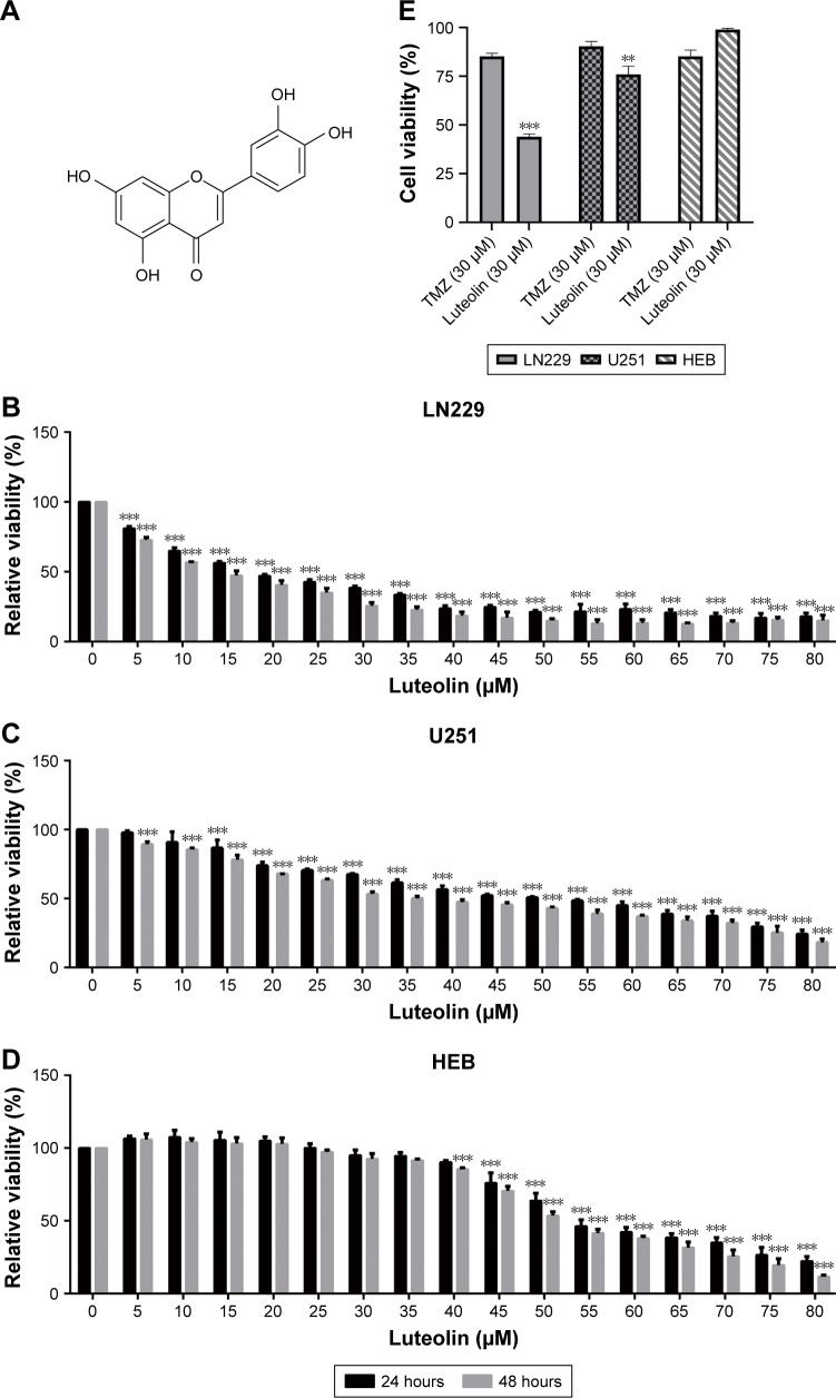
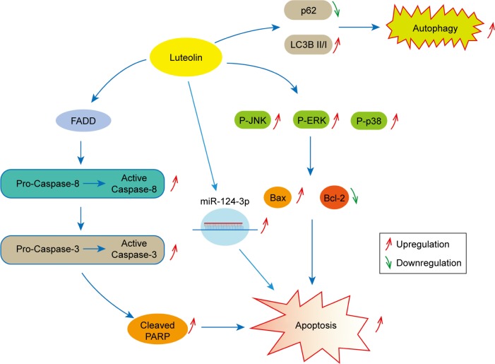
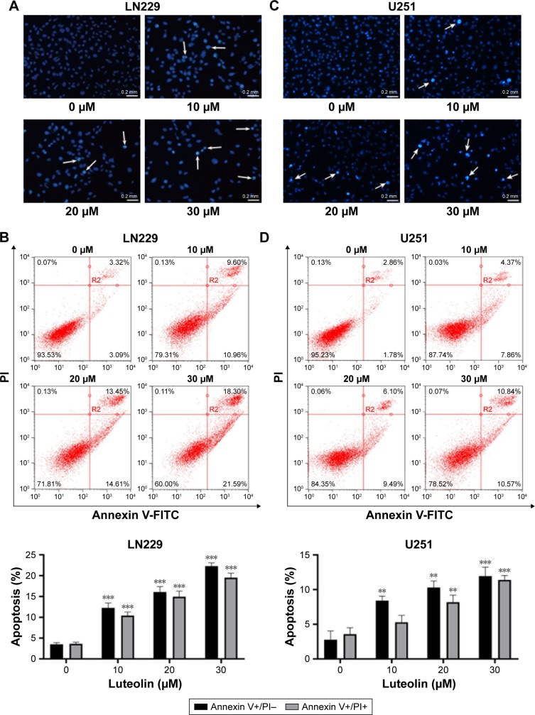
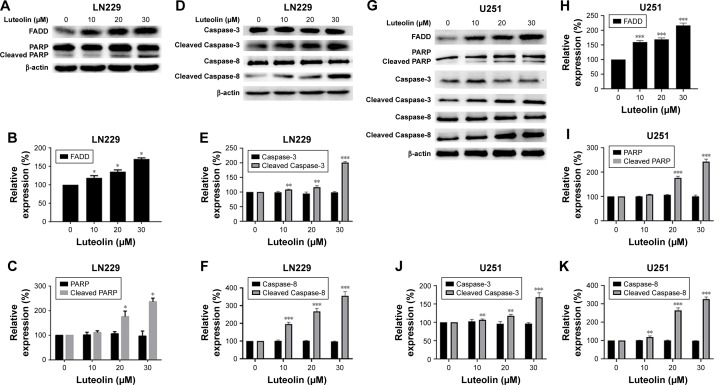
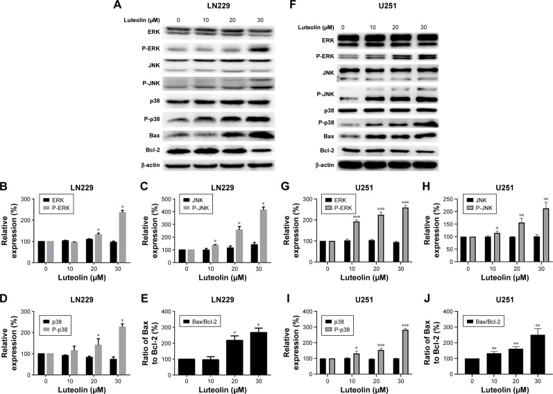
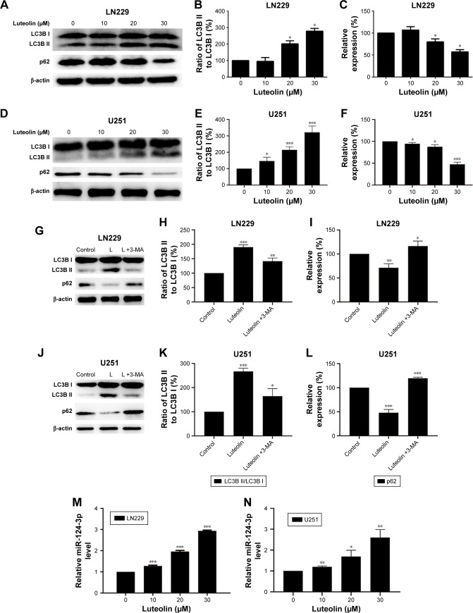
CCK-8 assays indicated that luteolin significantly inhibited glioma cell proliferation in a time- and dose-dependent manner. Fluorescent microscopy and flow cytometry analysis confirmed that luteolin induced glioma cell apoptosis. Western blot analysis showed that luteolin induced cellular apoptosis in glioma cells via MAPK activation (JNK, ERK, and p38). Luteolin stimulated the death receptor (FADD) to regulate the apoptosis proteins (Caspase-8, Caspase-3, and PARP). Luteolin increased the expression levels of LC3B II/I and downregulated the level of p62 that promotes cell autophagy. Finally, qRT-PCR confirmed that luteolin upregulated the expression levels of miR-124-3p.
These findings illustrate that luteolin may be a potential drug for glioma treatment.
胶质瘤是一种起源于脑和脊髓的恶性肿瘤,难以完全切除。尽管胶质瘤患者接受了积极治疗,但其生存率仍然很低。因此,迫切需要发现一种新的药物来治疗胶质瘤患者以提高生存率。在本研究中,我们在体外探索了木犀草素对胶质瘤的抗癌作用及其潜在机制。
采用细胞计数试剂盒-8 (CCK-8) 法测定细胞活力。利用荧光显微镜和流式细胞术分析来确定细胞凋亡情况。进行蛋白质印迹分析以探索蛋白质表达的变化。利用定量逆转录聚合酶链反应(qRT-PCR)分析来评估肿瘤抑制因子miR-124-3p的表达水平。
CCK-8试验表明,木犀草素以时间和剂量依赖性方式显著抑制胶质瘤细胞增殖。荧光显微镜和流式细胞术分析证实木犀草素诱导胶质瘤细胞凋亡。蛋白质印迹分析表明,木犀草素通过激活丝裂原活化蛋白激酶(JNK、ERK和p38)诱导胶质瘤细胞凋亡。木犀草素刺激死亡受体(FADD)来调节凋亡蛋白(半胱天冬酶-8、半胱天冬酶-3和聚(ADP-核糖)聚合酶)。木犀草素增加了LC3B II/I的表达水平,并下调了促进细胞自噬的p62水平。最后,qRT-PCR证实木犀草素上调了miR-124-3p的表达水平。
这些发现表明木犀草素可能是一种治疗胶质瘤的潜在药物。