• 文献检索
  • 文档翻译
  • 深度研究
  • 学术资讯
  • Suppr Zotero 插件Zotero 插件
  • 邀请有礼
  • 套餐&价格
  • 历史记录
应用&插件
Suppr Zotero 插件Zotero 插件浏览器插件Mac 客户端Windows 客户端微信小程序
定价
高级版会员购买积分包购买API积分包
服务
文献检索文档翻译深度研究API 文档MCP 服务
关于我们
关于 Suppr公司介绍联系我们用户协议隐私条款
关注我们

Suppr 超能文献

核心技术专利:CN118964589B侵权必究
粤ICP备2023148730 号-1Suppr @ 2026

文献检索

告别复杂PubMed语法,用中文像聊天一样搜索,搜遍4000万医学文献。AI智能推荐,让科研检索更轻松。

立即免费搜索

文件翻译

保留排版,准确专业,支持PDF/Word/PPT等文件格式,支持 12+语言互译。

免费翻译文档

深度研究

AI帮你快速写综述,25分钟生成高质量综述,智能提取关键信息,辅助科研写作。

立即免费体验

施莱姆氏管内皮细胞连接在巨囊泡形成中的作用:三维电子显微镜研究。

The Role of Schlemm's Canal Endothelium Cellular Connectivity in Giant Vacuole Formation: A 3D Electron Microscopy Study.

机构信息

Department of Ophthalmology, Boston University School of Medicine, Boston, Massachusetts, United States.

The Affiliated Eye Hospital of Wenzhou Medical University, Wenzhou, Zhejiang, China.

出版信息

Invest Ophthalmol Vis Sci. 2019 Apr 1;60(5):1630-1643. doi: 10.1167/iovs.18-26011.

DOI:10.1167/iovs.18-26011
PMID:30995299
原文链接:https://pmc.ncbi.nlm.nih.gov/articles/PMC6736380/
Abstract

PURPOSE

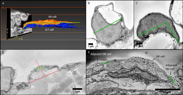
We investigated whether cellular connectivity between Schlemm's canal (SC) inner wall (IW) endothelium, and juxtacanalicular connective tissue (JCT), and between IW endothelial cells, plays a role in giant vacuole (GV) and pore formation by comparing perfusion- and immersion-fixed eyes.

METHODS

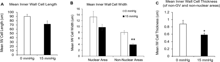
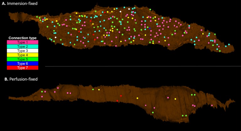
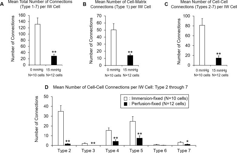
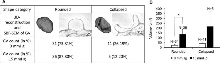
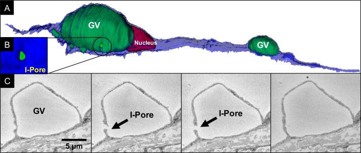
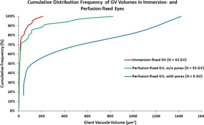
Normal human donor eyes (n = 4) were either immersion-fixed (0 mm Hg) or perfusion-fixed (15 mm Hg). Trabecular meshwork near SC was imaged using serial block-face scanning electron microscopy. A total of 12 IW cells from each group were 3D-reconstructed from ∼7040 electron micrographs and compared. In each cell, connections between IW cells and JCT cells/matrix were quantified; IW/IW connectivity was measured by cell border overlap length. GV volume, density, shape, and intracellular and paracellular pores were analyzed.

RESULTS

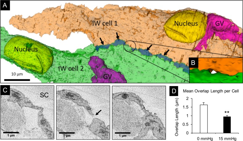
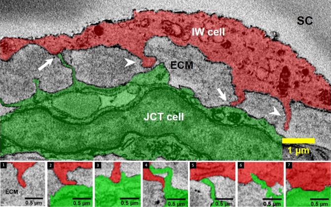
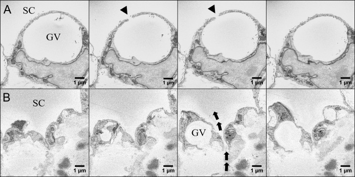
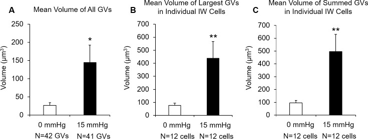
The mean number of IW/JCT cell-cell connections per cell significantly decreased (P < 0.01) while the summed GV volume per cell significantly increased (P < 0.01) in perfusion-fixed eyes compared to immersion-fixed eyes. Intracellular pores were observed in 14.6% of GVs in perfusion-fixed eyes and not observed in immersion-fixed eyes. The mean IW/IW overlap length per cell decreased (P < 0.01), and paracellular pores were found only in regions where IW/IW connectivity was minimal (overlap length = 0 μm) in perfusion-fixed eyes and not observed in immersion-fixed eyes.

CONCLUSIONS

Our data suggest that changes in IW/JCT connectivity may be an important factor in the formation of larger GVs, and decreased IW/IW connectivity may promote paracellular pore formation. Targeting the IW/JCT and IW/IW connectivity may therefore be a potential strategy to regulate outflow resistance and IOP. .

摘要

目的

通过比较灌注固定和浸泡固定的眼睛,我们研究了施莱姆管(SC)内皮层(IW)内皮细胞与小梁网间隙连接组织(JCT)之间以及 IW 内皮细胞之间的细胞连接是否在巨泡(GV)和孔形成中起作用。

方法

正常的人供体眼球(n = 4)分别进行浸泡固定(0 毫米汞柱)或灌注固定(15 毫米汞柱)。使用连续块面扫描电子显微镜对 SC 附近的小梁网进行成像。从每组中总共重建了 12 个 IW 细胞,从大约 7040 张电子显微镜照片中进行了比较。在每个细胞中,IW 细胞与 JCT 细胞/基质之间的连接数量进行了量化;通过细胞边界重叠长度测量 IW/IW 连接性。分析了 GV 体积、密度、形状以及细胞内和细胞旁孔。

结果

与浸泡固定的眼睛相比,灌注固定的眼睛中每个细胞的 IW/JCT 细胞-细胞连接数量显著减少(P < 0.01),而每个细胞的 GV 体积总和显著增加(P < 0.01)。在灌注固定的眼睛中观察到 14.6%的 GV 中存在细胞内孔,而在浸泡固定的眼睛中未观察到。每个细胞的 IW/IW 重叠长度平均值减少(P < 0.01),并且仅在灌注固定的眼睛中 IW/IW 连接性最小(重叠长度= 0 μm)的区域中发现细胞旁孔,而在浸泡固定的眼睛中未观察到。

结论

我们的数据表明,IW/JCT 连接性的变化可能是形成更大 GV 的一个重要因素,而 IW/IW 连接性的降低可能会促进细胞旁孔的形成。因此,靶向 IW/JCT 和 IW/IW 连接性可能是调节流出阻力和 IOP 的潜在策略。

https://cdn.ncbi.nlm.nih.gov/pmc/blobs/ea5c/6736380/7c85a7a2b143/i1552-5783-60-5-1630-f11.jpg
https://cdn.ncbi.nlm.nih.gov/pmc/blobs/ea5c/6736380/0e38866aa606/i1552-5783-60-5-1630-f01.jpg
https://cdn.ncbi.nlm.nih.gov/pmc/blobs/ea5c/6736380/d4f8cc66a40e/i1552-5783-60-5-1630-f02.jpg
https://cdn.ncbi.nlm.nih.gov/pmc/blobs/ea5c/6736380/9157eaf99f53/i1552-5783-60-5-1630-f03.jpg
https://cdn.ncbi.nlm.nih.gov/pmc/blobs/ea5c/6736380/5fe7ad06bbf0/i1552-5783-60-5-1630-f04.jpg
https://cdn.ncbi.nlm.nih.gov/pmc/blobs/ea5c/6736380/3ff25c4a0474/i1552-5783-60-5-1630-f05.jpg
https://cdn.ncbi.nlm.nih.gov/pmc/blobs/ea5c/6736380/584e7248fc1a/i1552-5783-60-5-1630-f06.jpg
https://cdn.ncbi.nlm.nih.gov/pmc/blobs/ea5c/6736380/2f617e429b09/i1552-5783-60-5-1630-f07.jpg
https://cdn.ncbi.nlm.nih.gov/pmc/blobs/ea5c/6736380/47221f2092fa/i1552-5783-60-5-1630-f08.jpg
https://cdn.ncbi.nlm.nih.gov/pmc/blobs/ea5c/6736380/7d4791b7bf2b/i1552-5783-60-5-1630-f09.jpg
https://cdn.ncbi.nlm.nih.gov/pmc/blobs/ea5c/6736380/dc026c465dc5/i1552-5783-60-5-1630-f10.jpg
https://cdn.ncbi.nlm.nih.gov/pmc/blobs/ea5c/6736380/7c85a7a2b143/i1552-5783-60-5-1630-f11.jpg

相似文献

1
The Role of Schlemm's Canal Endothelium Cellular Connectivity in Giant Vacuole Formation: A 3D Electron Microscopy Study.施莱姆氏管内皮细胞连接在巨囊泡形成中的作用:三维电子显微镜研究。
Invest Ophthalmol Vis Sci. 2019 Apr 1;60(5):1630-1643. doi: 10.1167/iovs.18-26011.
2
Morphological factors associated with giant vacuoles with I-pores in Schlemm's canal endothelial cells of human eyes: A serial block-face scanning electron microscopy study.人眼房水流出通道 Schlemm 管内皮细胞中具有 I 型孔的巨大空泡的形态学相关因素:一个连续块面扫描电子显微镜研究。
Exp Eye Res. 2021 Apr;205:108488. doi: 10.1016/j.exer.2021.108488. Epub 2021 Feb 9.
3
Schlemm's Canal Endothelium Cellular Connectivity in Giant Vacuole and Pore Formation in Different Flow-type Areas: A Serial Block-Face Scanning Electron Microscopy Study.施莱姆管内皮细胞在不同血流类型区域的巨大液泡和孔隙形成中的细胞连接性:一项连续块面扫描电子显微镜研究
Front Cell Dev Biol. 2022 Apr 13;10:867376. doi: 10.3389/fcell.2022.867376. eCollection 2022.
4
Colocalization of outflow segmentation and pores along the inner wall of Schlemm's canal.Schlemm管内壁上流出物分割与孔隙的共定位。
Exp Eye Res. 2015 Jan;130:87-96. doi: 10.1016/j.exer.2014.11.008. Epub 2014 Nov 13.
5
Interendothelial junctions in normal human Schlemm's canal respond to changes in pressure.正常人类小梁网中的内皮细胞间连接对压力变化有反应。
Invest Ophthalmol Vis Sci. 1997 Nov;38(12):2460-8.
6
Comparative studies between species that do and do not exhibit the washout effect.对表现出和未表现出洗脱效应的物种之间的比较研究。
Exp Eye Res. 2007 Mar;84(3):435-43. doi: 10.1016/j.exer.2006.10.015. Epub 2006 Dec 13.
7
Effect of fixation pressure on juxtacanalicular tissue and Schlemm's canal.固定压力对近小管组织和施莱姆管的影响。
Invest Ophthalmol Vis Sci. 1996 Jan;37(1):114-24.
8
The mechanism of increasing outflow facility by rho-kinase inhibition with Y-27632 in bovine eyes.Y-27632通过抑制牛眼的rho激酶增加房水流出易度的机制。
Exp Eye Res. 2008 Feb;86(2):271-81. doi: 10.1016/j.exer.2007.10.018. Epub 2007 Nov 5.
9
Similar hydrodynamic and morphological changes in the aqueous humor outflow pathway after washout and Y27632 treatment in monkey eyes.猴眼灌洗和 Y27632 处理后房水流出通路中相似的流体动力学和形态学变化。
Exp Eye Res. 2011 Oct;93(4):397-404. doi: 10.1016/j.exer.2011.05.012. Epub 2011 Jun 6.
10
The relationship between pore density and outflow facility in human eyes.人眼的孔隙密度与房水流畅系数之间的关系。
Invest Ophthalmol Vis Sci. 1992 Apr;33(5):1661-9.

引用本文的文献

1
Investigation of two pore types in Schlemm's canal inner wall endothelium in flow-type areas using serial block-face scanning electron microscopy.使用连续块面扫描电子显微镜对施莱姆管内壁内皮细胞中流动型区域的两种孔隙类型进行研究。
Exp Eye Res. 2025 Oct;259:110577. doi: 10.1016/j.exer.2025.110577. Epub 2025 Aug 11.
2
Mechanobiology in the eye.眼部的力学生物学
NPJ Biol Phys Mech. 2025;2(1):18. doi: 10.1038/s44341-025-00022-6. Epub 2025 Jul 4.
3
3D traction force microscopy in human trabecular meshwork tissues: Effects of ROCK and YAP/TAZ inhibition in normal and glaucomatous tissues.

本文引用的文献

1
Netarsudil Increases Outflow Facility in Human Eyes Through Multiple Mechanisms.奈他地尔通过多种机制增加人眼的房水流出率。
Invest Ophthalmol Vis Sci. 2016 Nov 1;57(14):6197-6209. doi: 10.1167/iovs.16-20189.
2
Variations in active outflow along the trabecular outflow pathway.小梁网流出途径中有效房水流出的差异。
Exp Eye Res. 2016 May;146:354-360. doi: 10.1016/j.exer.2016.01.008. Epub 2016 Jan 13.
3
The 3D pore structure and fluid dynamics simulation of macroporous monoliths: High permeability due to alternating channel width.
人小梁网组织中的三维牵引力显微镜检查:ROCK和YAP/TAZ抑制对正常和青光眼组织的影响。
Tissue Cell. 2025 Jun 6;96:103005. doi: 10.1016/j.tice.2025.103005.
4
Endothelial glycocalyx in different flow regions of the trabecular outflow pathway in bovine eyes.牛眼小梁网流出途径不同血流区域的内皮糖萼。
Front Cell Dev Biol. 2025 Apr 25;13:1569569. doi: 10.3389/fcell.2025.1569569. eCollection 2025.
5
High-resolution modeling of aqueous humor dynamics in the conventional outflow pathway of a normal human donor eye.正常人类供体眼传统流出途径中房水动力学的高分辨率建模。
Comput Methods Programs Biomed. 2025 Mar;260:108538. doi: 10.1016/j.cmpb.2024.108538. Epub 2024 Nov 29.
6
ECM biomaterials for modeling of outflow cell biology in health and disease.用于健康和疾病状态下流出细胞生物学建模的细胞外基质生物材料
Biomater Biosyst. 2024 Mar 5;13:100091. doi: 10.1016/j.bbiosy.2024.100091. eCollection 2024 Mar.
7
Implementing new computational methods for the study of JCT and SC inner wall basement membrane biomechanics and hydrodynamics.研究 JCT 和 SC 内皮层基底膜生物力学和流体动力学的新计算方法的实施。
Comput Methods Programs Biomed. 2024 Jan;243:107909. doi: 10.1016/j.cmpb.2023.107909. Epub 2023 Oct 31.
8
Biomechanics of the JCT and SC Inner Wall Endothelial Cells with Their Basement Membrane Using 3D Serial Block-Face Scanning Electron Microscopy.使用三维连续块面扫描电子显微镜对关节软骨下骨和滑膜衬里内层内皮细胞及其基底膜的生物力学研究
Bioengineering (Basel). 2023 Sep 4;10(9):1038. doi: 10.3390/bioengineering10091038.
9
Intracameral Injection of AAV-DJ.COMP-ANG1 Reduces the IOP of Mice by Reshaping the Trabecular Outflow Pathway.房水内注射 AAV-DJ.COMP-ANG1 通过重塑小梁流出通路降低小鼠眼压。
Invest Ophthalmol Vis Sci. 2022 Dec 1;63(13):15. doi: 10.1167/iovs.63.13.15.
10
Schlemm's Canal Endothelium Cellular Connectivity in Giant Vacuole and Pore Formation in Different Flow-type Areas: A Serial Block-Face Scanning Electron Microscopy Study.施莱姆管内皮细胞在不同血流类型区域的巨大液泡和孔隙形成中的细胞连接性:一项连续块面扫描电子显微镜研究
Front Cell Dev Biol. 2022 Apr 13;10:867376. doi: 10.3389/fcell.2022.867376. eCollection 2022.
大孔整体材料的三维孔隙结构与流体动力学模拟:通道宽度交替变化导致高渗透性
J Chromatogr A. 2015 Dec 18;1425:141-9. doi: 10.1016/j.chroma.2015.11.026. Epub 2015 Nov 14.
4
Multiple Retinal Axons Converge onto Relay Cells in the Adult Mouse Thalamus.多条视网膜轴突汇聚到成年小鼠丘脑的中继细胞上。
Cell Rep. 2015 Sep 8;12(10):1575-83. doi: 10.1016/j.celrep.2015.08.003. Epub 2015 Aug 28.
5
Automated tracing of myelinated axons and detection of the nodes of Ranvier in serial images of peripheral nerves.外周神经连续图像中髓鞘化轴突的自动追踪及郎飞结的检测
J Microsc. 2015 Aug;259(2):143-154. doi: 10.1111/jmi.12266. Epub 2015 Jun 12.
6
Serial block face-scanning electron microscopy: a method to study retinal degenerative phenotypes.连续块面扫描电子显微镜术:一种研究视网膜退行性表型的方法。
Curr Protoc Mouse Biol. 2014 Dec 11;4(4):197-204. doi: 10.1002/9780470942390.mo140169.
7
Colocalization of outflow segmentation and pores along the inner wall of Schlemm's canal.Schlemm管内壁上流出物分割与孔隙的共定位。
Exp Eye Res. 2015 Jan;130:87-96. doi: 10.1016/j.exer.2014.11.008. Epub 2014 Nov 13.
8
Biomechanical strain as a trigger for pore formation in Schlemm's canal endothelial cells.生物力学应变作为施莱姆管内皮细胞中孔隙形成的触发因素。
Exp Eye Res. 2014 Oct;127:224-35. doi: 10.1016/j.exer.2014.08.003. Epub 2014 Aug 14.
9
Morphological and hydrodynamic correlations with increasing outflow facility by rho-kinase inhibitor Y-27632.通过 rho 激酶抑制剂 Y-27632 增加流出道顺应性的形态学和流体动力学相关性。
J Ocul Pharmacol Ther. 2014 Mar-Apr;30(2-3):143-53. doi: 10.1089/jop.2013.0192. Epub 2014 Jan 24.
10
Multi-scale analysis of segmental outflow patterns in human trabecular meshwork with changing intraocular pressure.多尺度分析眼压变化时人眼小梁网节段性流出模式
J Ocul Pharmacol Ther. 2014 Mar-Apr;30(2-3):213-23. doi: 10.1089/jop.2013.0182. Epub 2014 Jan 23.