The First Department of Gynecologic Oncology and Gynecology, Medical University of Lublin, Lublin, Poland.
Tumor Immunology Laboratory, Medical University of Lublin, Lublin, Poland.
Front Immunol. 2019 Apr 3;10:691. doi: 10.3389/fimmu.2019.00691. eCollection 2019.
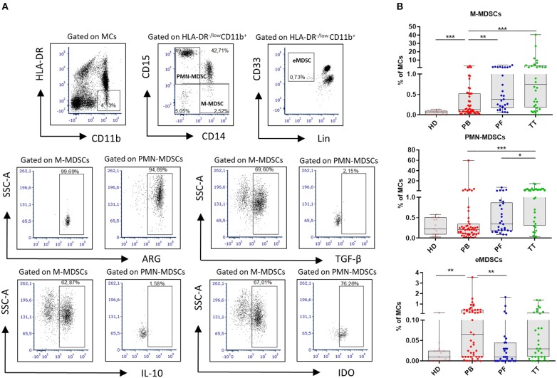
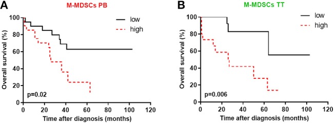
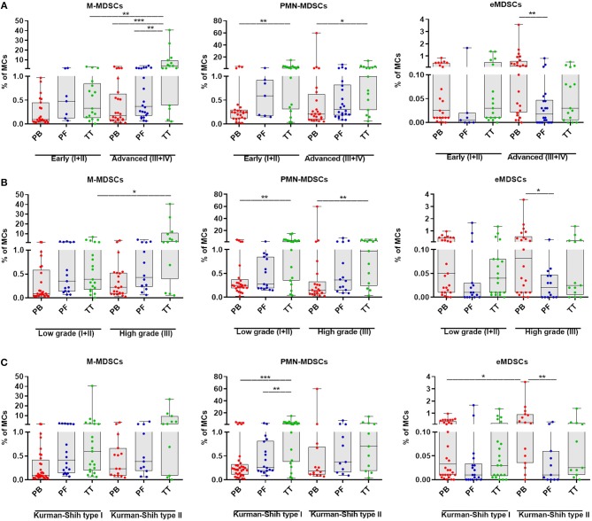
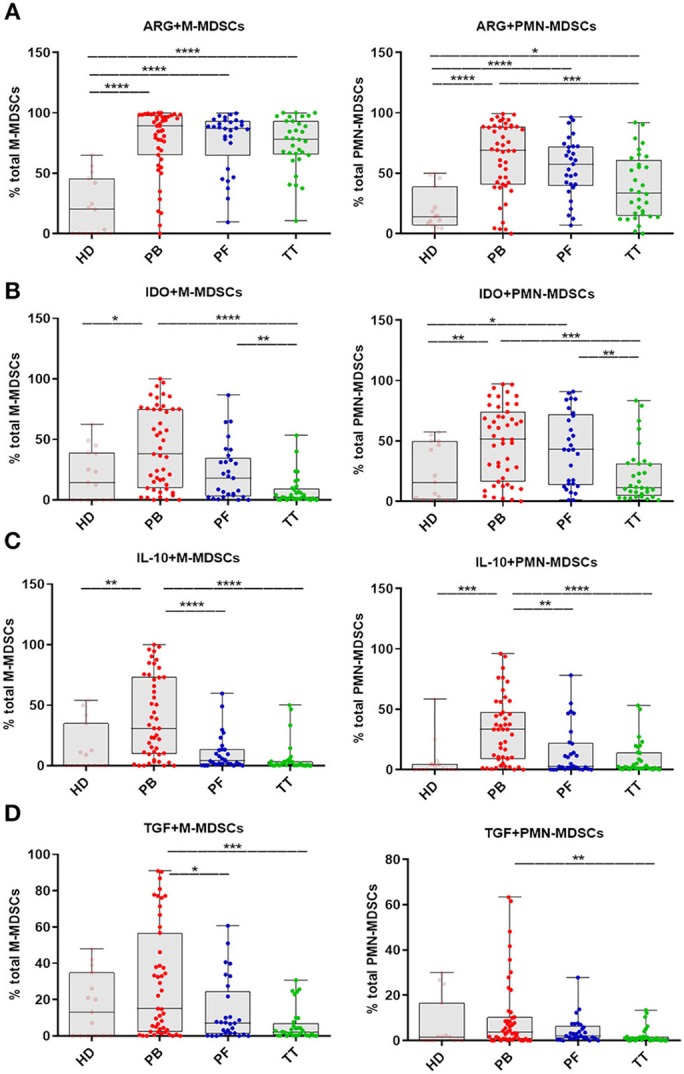
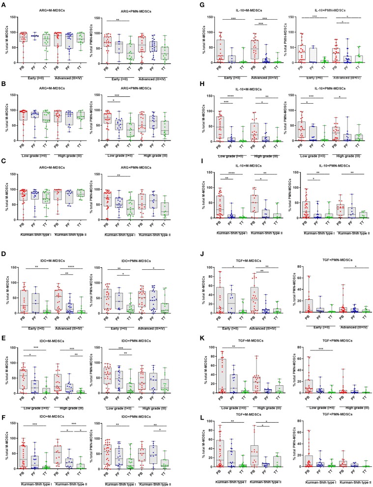
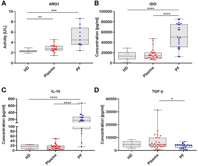
Myeloid-derived suppressor cells (MDSCs) expansion is a hallmark of cancer. Three major MDSC subsets defined as monocytic (M)-MDSCs, polymorphonuclear (PMN)-MDSCs and early stage (e)MDSCs can be revealed in human diseases. However, the clinical relevance and immunosupressive pattern of these cells in epithelial ovarian cancer (EOC) are unknown. Therefore, we performed a comprehensive analysis of each MDSC subset and immunosupressive factors in the peripheral blood (PB), peritoneal fluid (PF), and the tumor tissue (TT) samples from EOC and integrated this data with the patients' clinicopathological characteristic. MDSCs were analyzed using multicolor flow cytometry. Immunosuppressive factors analysis was performed with ELISA and qRT-PCR. The level of M-MDSCs in the PB/PF/TT of EOC was significantly higher than in healthy donors (HD); frequency of PMN-MDSCs was significantly greater in the TT than in the PB/PF and HD; while the level of eMDSCs was greater in the PB compared with the PF and HD. Elevated abundance of tumor-infiltrating M-MDSCs was associated with advanced stage and high grade of EOC. An analysis of immunosuppressive pattern showed significantly increased blood-circulating ARG/IDO/IL-10-expressing M- and PMN-MDSCs in the EOC patients compared with HD and differences in the accumulation of these subsets in the three tumor immune microenvironments (TIME). This accumulation was positively correlated with levels of TGF-β and ARG1 in the plasma and PF. Low level of blood-circulating and tumor-infiltrating M-MDSCs, but neither PMN-MDSCs nor eMDSCs was strongly associated with prolonged survival in ovarian cancer patients. Our results highlight M-MDSCs as the subset with potential the highest clinical significance.
髓系来源的抑制细胞(MDSCs)扩增是癌症的一个标志。在人类疾病中,可以发现三种主要的 MDSC 亚群,分别为单核细胞(M)-MDSCs、多形核(PMN)-MDSCs 和早期(e)MDSCs。然而,这些细胞在卵巢上皮癌(EOC)中的临床相关性和免疫抑制模式尚不清楚。因此,我们对来自 EOC 的外周血(PB)、腹腔液(PF)和肿瘤组织(TT)样本中的每个 MDSC 亚群和免疫抑制因子进行了全面分析,并将这些数据与患者的临床病理特征进行了整合。使用多色流式细胞术分析 MDSCs。通过 ELISA 和 qRT-PCR 分析免疫抑制因子。EOC 的 PB/PF/TT 中 M-MDSCs 的水平明显高于健康供体(HD);TT 中 PMN-MDSCs 的频率明显高于 PB/PF 和 HD;而 eMDSCs 的水平在 PB 中高于 PF 和 HD。肿瘤浸润性 M-MDSCs 的丰度升高与 EOC 的晚期和高级别相关。免疫抑制模式分析显示,与 HD 相比,EOC 患者血液中循环的 ARG/IDO/IL-10 表达的 M 和 PMN-MDSCs 明显增加,并且这两种亚群在三种肿瘤免疫微环境(TIME)中的积累存在差异。这种积累与血浆和 PF 中 TGF-β和 ARG1 的水平呈正相关。血液和肿瘤浸润性 M-MDSCs 水平低,PMN-MDSCs 和 eMDSCs 水平低与卵巢癌患者的生存时间延长强烈相关。我们的研究结果突出了 M-MDSCs 作为具有最高临床意义的亚群。