• 文献检索
  • 文档翻译
  • 深度研究
  • 学术资讯
  • Suppr Zotero 插件Zotero 插件
  • 邀请有礼
  • 套餐&价格
  • 历史记录
应用&插件
Suppr Zotero 插件Zotero 插件浏览器插件Mac 客户端Windows 客户端微信小程序
定价
高级版会员购买积分包购买API积分包
服务
文献检索文档翻译深度研究API 文档MCP 服务
关于我们
关于 Suppr公司介绍联系我们用户协议隐私条款
关注我们

Suppr 超能文献

核心技术专利:CN118964589B侵权必究
粤ICP备2023148730 号-1Suppr @ 2026

文献检索

告别复杂PubMed语法,用中文像聊天一样搜索,搜遍4000万医学文献。AI智能推荐,让科研检索更轻松。

立即免费搜索

文件翻译

保留排版,准确专业,支持PDF/Word/PPT等文件格式,支持 12+语言互译。

免费翻译文档

深度研究

AI帮你快速写综述,25分钟生成高质量综述,智能提取关键信息,辅助科研写作。

立即免费体验

伴有出血性心包积液的右心移行性肿块:诊断难题

Right Heart Mass in Transit with a Hemorrhagic Pericardial Effusion: A Diagnostic Dilemma.

作者信息

Nanda Amit, Khouzam Rami N, Jefferies John, Moon Marc, Makan Majesh

机构信息

Internal Medicine, University of Tennessee, Memphis, USA.

Cardiology, University of Tennessee, Memphis, USA.

出版信息

Cureus. 2019 Feb 4;11(2):e4009. doi: 10.7759/cureus.4009.

DOI:10.7759/cureus.4009
PMID:31001463
原文链接:https://pmc.ncbi.nlm.nih.gov/articles/PMC6450589/
Abstract

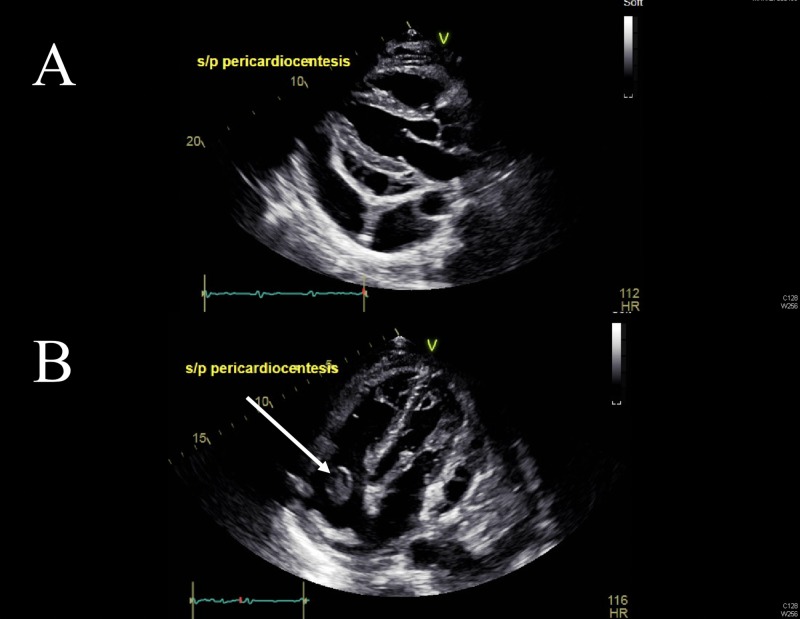
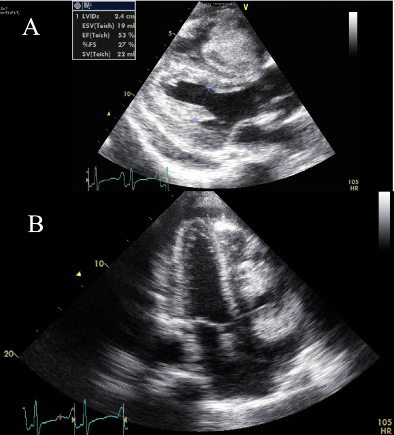
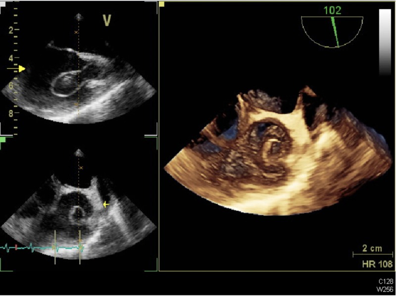
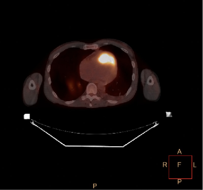
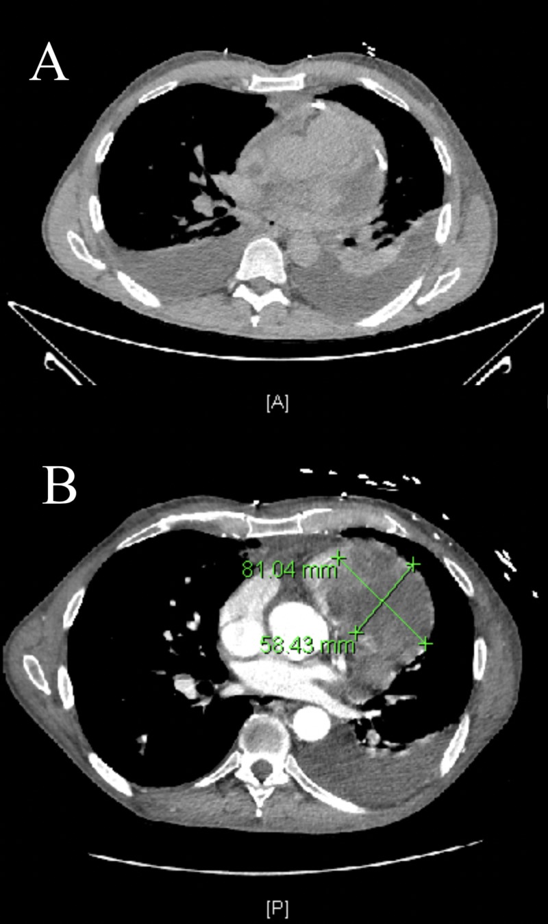
Metastatic disease to the heart is more often a post-mortem diagnosis due to non-specific symptoms and a low index of suspicion. Our case is a unique presentation of a rare case of cardiac metastasis from oropharyngeal cancer, which eluded echocardiographic diagnosis despite the presence of a hemorrhagic pericardial effusion. The cardiac metastasis, in fact, starts as pericardial seeding, as illustrated by the positron emission tomography (PET) imaging. The pericardial metastatic disease then becomes rapidly invasive into the cardiac chambers, hence presenting as a large mass on the echocardiogram and computed tomography (CT) scan of the chest. This is the first such case of pericardial metastasis from a squamous cell carcinoma of the tongue being reported and highlights the importance of an aggressive multimodality diagnostic approach in cases where such a clinical suspicion exists. While a two-dimensional (2D) echocardiogram is the most readily available modality, we recommend that this is complemented by the use of a three-dimensional (3D) echocardiogram, as well as metabolic and radiologic imaging with PET and CT scans.

摘要

由于症状不具特异性且怀疑指数较低,心脏转移性疾病往往是尸检诊断。我们的病例是口咽癌心脏转移罕见病例的独特呈现,尽管存在出血性心包积液,但超声心动图仍未能诊断出来。事实上,正电子发射断层扫描(PET)成像显示,心脏转移始于心包播散。心包转移性疾病随后迅速侵入心腔,因此在超声心动图和胸部计算机断层扫描(CT)上表现为巨大肿块。这是首次报道舌鳞状细胞癌心包转移的此类病例,强调了在存在此类临床怀疑的病例中采用积极的多模态诊断方法的重要性。虽然二维(2D)超声心动图是最容易获得的检查方式,但我们建议使用三维(3D)超声心动图以及PET和CT扫描的代谢和放射成像作为补充。

https://cdn.ncbi.nlm.nih.gov/pmc/blobs/8c94/6450589/634d305dd95c/cureus-0011-00000004009-i05.jpg
https://cdn.ncbi.nlm.nih.gov/pmc/blobs/8c94/6450589/73a45fbbd895/cureus-0011-00000004009-i01.jpg
https://cdn.ncbi.nlm.nih.gov/pmc/blobs/8c94/6450589/1285e0383063/cureus-0011-00000004009-i02.jpg
https://cdn.ncbi.nlm.nih.gov/pmc/blobs/8c94/6450589/836e4829f793/cureus-0011-00000004009-i03.jpg
https://cdn.ncbi.nlm.nih.gov/pmc/blobs/8c94/6450589/3159c696198a/cureus-0011-00000004009-i04.jpg
https://cdn.ncbi.nlm.nih.gov/pmc/blobs/8c94/6450589/634d305dd95c/cureus-0011-00000004009-i05.jpg

相似文献

1
Right Heart Mass in Transit with a Hemorrhagic Pericardial Effusion: A Diagnostic Dilemma.伴有出血性心包积液的右心移行性肿块:诊断难题
Cureus. 2019 Feb 4;11(2):e4009. doi: 10.7759/cureus.4009.
2
A Case of Low-Grade Primary Cardiac Lymphoma with Pericardial Effusion Diagnosed by Combined 18F-Fluorodeoxyglucose Positron Emission Tomography and Computed Tomography (FDG-PET/CT) Imaging and Effusion Cytology.一例经18F-氟脱氧葡萄糖正电子发射断层扫描与计算机断层扫描(FDG-PET/CT)成像及积液细胞学检查诊断的伴有心包积液的原发性低度心脏淋巴瘤病例
Am J Case Rep. 2018 Mar 14;19:292-295. doi: 10.12659/ajcr.908385.
3
More advantages in detecting bone and soft tissue metastases from prostate cancer using F-PSMA PET/CT.使用F-PSMA PET/CT检测前列腺癌骨和软组织转移方面有更多优势。
Hell J Nucl Med. 2019 Jan-Apr;22(1):6-9. doi: 10.1967/s002449910952. Epub 2019 Mar 7.
4
The role of early contrast-enhanced chest computed tomography in the aetiological diagnosis of patients presenting with cardiac tamponade or large pericardial effusion.早期对比增强胸部计算机断层扫描在心脏压塞或大量心包积液患者病因诊断中的作用。
Eur Heart J Cardiovasc Imaging. 2016 Apr;17(4):421-8. doi: 10.1093/ehjci/jev225. Epub 2015 Sep 15.
5
[Clinical and radiologic characteristics as well as outcomes of patients with primary cardiac angiosarcoma].[原发性心脏血管肉瘤患者的临床及影像学特征与预后]
Zhonghua Xin Xue Guan Bing Za Zhi. 2019 Sep 24;47(9):731-736. doi: 10.3760/cma.j.issn.0253-3758.2019.09.015.
6
Pericardial Metastasis From Recurrent Squamous Cell Carcinoma of Hypopharynx in a Living Patient.一名在世患者下咽复发性鳞状细胞癌的心包转移
Cureus. 2022 Apr 17;14(4):e24216. doi: 10.7759/cureus.24216. eCollection 2022 Apr.
7
European Association of Cardiovascular Imaging/Cardiovascular Imaging Department of the Brazilian Society of Cardiology recommendations for the use of cardiac imaging to assess and follow patients after heart transplantation.欧洲心血管影像协会/巴西心脏病学会心血管影像学部关于心脏移植后评估和随访患者使用心脏影像的建议。
Eur Heart J Cardiovasc Imaging. 2015 Sep;16(9):919-48. doi: 10.1093/ehjci/jev139. Epub 2015 Jul 2.
8
[Hemorrhagic nonspecific pericardial effusion as an initial symptom of angiosarcoma of the right heart].[出血性非特异性心包积液作为右心血管肉瘤的首发症状]
Z Kardiol. 2004 Oct;93(10):807-12. doi: 10.1007/s00392-004-0124-y.
9
Malignant pericardial effusion not diagnosed by pericardial fluid and biopsy: importance of CT scan.
Int J Cardiol. 2007 Apr 12;117(1):e53-5. doi: 10.1016/j.ijcard.2006.11.089. Epub 2007 Feb 9.
10
De-Differentiated Myxofibrosarcoma Metastasis in the Right Atrium: An Invasive But Diagnostic Approach.右心房去分化黏液纤维肉瘤转移:一种侵入性但具有诊断意义的方法。
JACC Case Rep. 2019 Aug 21;1(2):151-155. doi: 10.1016/j.jaccas.2019.06.021. eCollection 2019 Aug.

引用本文的文献

1
Systematic Review: Cardiac Metastasis of Lingual Squamous Cell Carcinoma.系统评价:舌鳞状细胞癌的心脏转移
Spartan Med Res J. 2021 Aug 30;6(2):27297. doi: 10.51894/001c.27297. eCollection 2021.

本文引用的文献

1
Integrated 18F-FDG PET/MR imaging in the assessment of cardiac masses: a pilot study.18F-FDG PET/MR 成像综合评估心脏肿物:一项初步研究
J Nucl Med. 2015 Feb;56(2):255-60. doi: 10.2967/jnumed.114.147744. Epub 2014 Dec 31.
2
MR imaging of cardiac tumors and masses: a review of methods and clinical applications.心脏肿瘤和肿块的磁共振成像:方法和临床应用综述。
Radiology. 2013 Jul;268(1):26-43. doi: 10.1148/radiol.13121239.
3
Early cardiac metastasis from squamous cell carcinoma of the tongue in 2 patients.2例舌鳞状细胞癌早期发生心脏转移。
Tex Heart Inst J. 2012;39(4):565-7.
4
Cardiac tumors in a tertiary care cancer hospital: clinical features, echocardiographic findings, treatment and outcomes.三级癌症专科医院的心脏肿瘤:临床特征、超声心动图表现、治疗及预后
Heart Int. 2012 Feb 3;7(1):e4. doi: 10.4081/hi.2012.e4. Epub 2012 Feb 13.
5
Differentiation of malignant and benign cardiac tumors using 18F-FDG PET/CT.应用 18F-FDG PET/CT 鉴别心脏良恶性肿瘤。
J Nucl Med. 2012 Jun;53(6):856-63. doi: 10.2967/jnumed.111.095364. Epub 2012 May 10.
6
Cardiac masses, part 2: key imaging features for diagnosis and surgical planning.心脏肿块,第 2 部分:诊断和手术规划的关键成像特征。
AJR Am J Roentgenol. 2011 Nov;197(5):W842-51. doi: 10.2214/AJR.11.6903.
7
How to differentiate benign versus malignant cardiac and paracardiac 18F FDG uptake at oncologic PET/CT.如何在肿瘤 PET/CT 上区分良性与恶性心脏和心脏旁 18F-FDG 摄取。
Radiographics. 2011 Sep-Oct;31(5):1287-305. doi: 10.1148/rg.315115003.
8
Cardiac metastasis of tongue cancer may cause sudden death.舌癌的心脏转移可能导致猝死。
Auris Nasus Larynx. 2008 Sep;35(3):423-5. doi: 10.1016/j.anl.2007.04.017. Epub 2007 Sep 14.
9
Cardiac metastases.心脏转移瘤。
J Clin Pathol. 2007 Jan;60(1):27-34. doi: 10.1136/jcp.2005.035105. Epub 2006 Nov 10.
10
Cardiac tumours: diagnosis and management.心脏肿瘤:诊断与管理
Lancet Oncol. 2005 Apr;6(4):219-28. doi: 10.1016/S1470-2045(05)70093-0.