Department of Translational Science & Molecular Medicine, College of Human Medicine, Michigan State University, Grand Rapids, MI, USA.
Hauenstein Neuroscience Center, Mercy Health Saint Mary's, Grand Rapids, Michigan, USA.
Mov Disord. 2019 May;34(5):697-707. doi: 10.1002/mds.27695. Epub 2019 Apr 19.
Levodopa-induced dyskinesias are an often debilitating side effect of levodopa therapy in Parkinson's disease. Although up to 90% of individuals with PD develop this side effect, uniformly effective and well-tolerated antidyskinetic treatment remains a significant unmet need. The pathognomonic loss of striatal dopamine in PD results in dysregulation and disinhibition of striatal CaV1.3 calcium channels, leading to synaptopathology that appears to be involved in levodopa-induced dyskinesias. Although there are clinically available drugs that can inhibit CaV1.3 channels, they are not adequately potent and have only partial and transient impact on levodopa-induced dyskinesias.
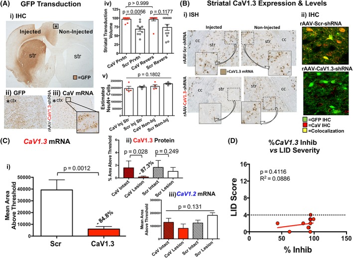
To provide unequivocal target validation, free of pharmacological limitations, we developed a CaV1.3 shRNA to provide high-potency, target-selective, mRNA-level silencing of striatal CaV1.3 channels and examined its ability to impact levodopa-induced dyskinesias in severely parkinsonian rats.
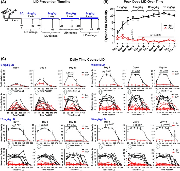
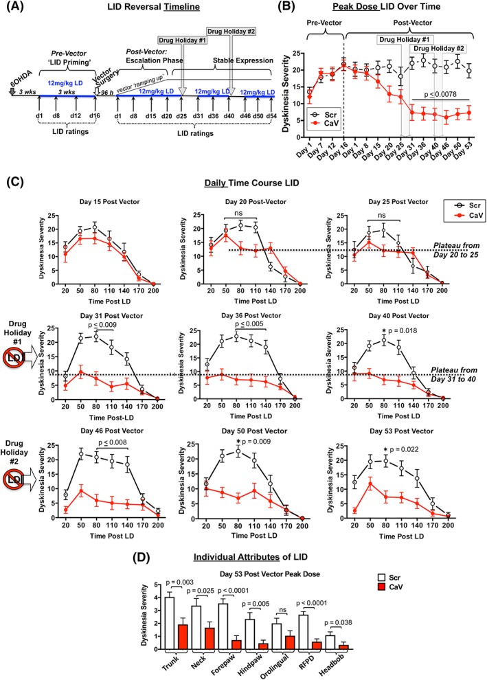
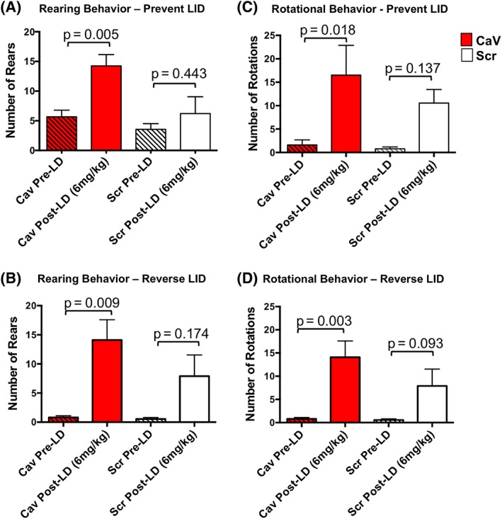
We demonstrate that vector-mediated silencing of striatal CaV1.3 expression in severely parkinsonian rats prior to the introduction of levodopa can uniformly and completely prevent induction of levodopa-induced dyskinesias, and this antidyskinetic benefit persists long term and with high-dose levodopa. In addition, this approach is capable of ameliorating preexisting severe levodopa-induced dyskinesias. Importantly, motoric responses to low-dose levodopa remained intact in the presence of striatal CaV1.3 silencing, indicating preservation of levodopa benefit without dyskinesia liability.
The current data provide some of the most profound antidyskinetic benefit reported to date and suggest that genetic silencing of striatal CaV1.3 channels has the potential to transform treatment of individuals with PD by allowing maintenance of motor benefit of levodopa in the absence of the debilitating levodopa-induced dyskinesia side effect. © 2019 The Authors. Movement Disorders published by Wiley Periodicals, Inc. on behalf of International Parkinson and Movement Disorder Society.
左旋多巴诱导的运动障碍是帕金森病患者左旋多巴治疗的一种常见且使人虚弱的副作用。尽管多达 90%的 PD 患者会出现这种副作用,但仍然需要一种统一有效且耐受性良好的抗运动障碍治疗方法。PD 中纹状体多巴胺的特征性丧失导致纹状体 CaV1.3 钙通道的失调和去抑制,导致突触病理学,这似乎与左旋多巴诱导的运动障碍有关。尽管有临床可用的药物可以抑制 CaV1.3 通道,但它们的效力不足,对左旋多巴诱导的运动障碍只有部分和短暂的影响。
为了提供明确的靶标验证,我们开发了一种 CaV1.3 shRNA,以提供纹状体 CaV1.3 通道的高效力、靶标选择性、mRNA 水平沉默,并用其来研究其在严重帕金森病大鼠中对左旋多巴诱导的运动障碍的影响。
我们证明,在引入左旋多巴之前,严重帕金森病大鼠的纹状体 CaV1.3 表达通过载体介导的沉默,可以均匀且完全地预防左旋多巴诱导的运动障碍的诱导,并且这种抗运动障碍的益处持久且在高剂量左旋多巴下仍然存在。此外,这种方法能够改善已存在的严重左旋多巴诱导的运动障碍。重要的是,在纹状体 CaV1.3 沉默的情况下,对低剂量左旋多巴的运动反应仍然完整,表明在没有运动障碍风险的情况下保留了左旋多巴的益处。
目前的数据提供了迄今为止报道的最显著的抗运动障碍益处,并表明纹状体 CaV1.3 通道的基因沉默有可能通过在不产生使人虚弱的左旋多巴诱导的运动障碍副作用的情况下维持左旋多巴的运动获益,从而改变 PD 患者的治疗方法。