Zhu Shuyi, Bergamino Maurizio, Fuentes Alberto, Sandoval Ivette M, Marmion David J, Bishop Christopher, Manfredsson Fredric P, Stokes Ashley M
Barrow Neuroimaging Innovation Center, Barrow Neurological Institute, Phoenix, AZ, United States.
School of Life Sciences, Arizona State University, Tempe, AZ, United States.
Front Neurosci. 2025 Jun 3;19:1591215. doi: 10.3389/fnins.2025.1591215. eCollection 2025.
Parkinson's Disease (PD), the second most common neurodegenerative disorder, is characterized by motor and non-motor symptoms linked to dopaminergic neuron degeneration. This study utilized the 6-hydroxydopamine (6-OHDA) rat model to replicate PD-like dopaminergic degeneration through targeted injections into the medial forebrain bundle and substantia nigra.
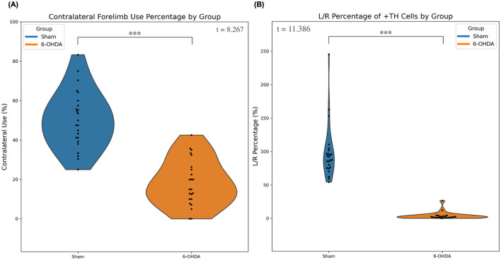
Behavioral assessments revealed hallmark motor deficits, while MRI was performed to assess complementary functional connectivity and structural connectivity. Post-mortem tyrosine hydroxylase (TH) staining confirmed extensive dopaminergic neuron loss, validating the pathological relevance of the model and ensuring data integrity. MRI data were collected at 7T in 46 male Fischer F344 rats (23 6-OHDA, 23 sham) to characterize functional and structural connectivity differences between cohorts.
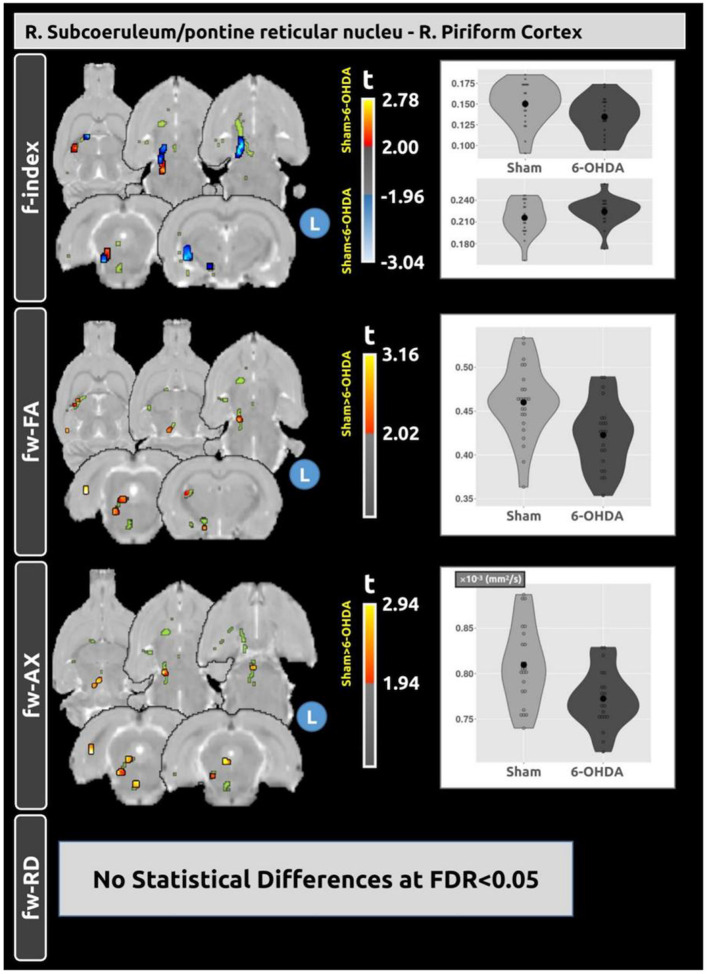
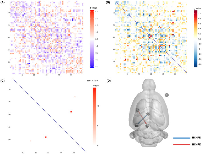
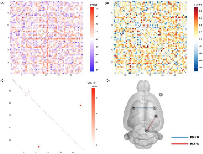
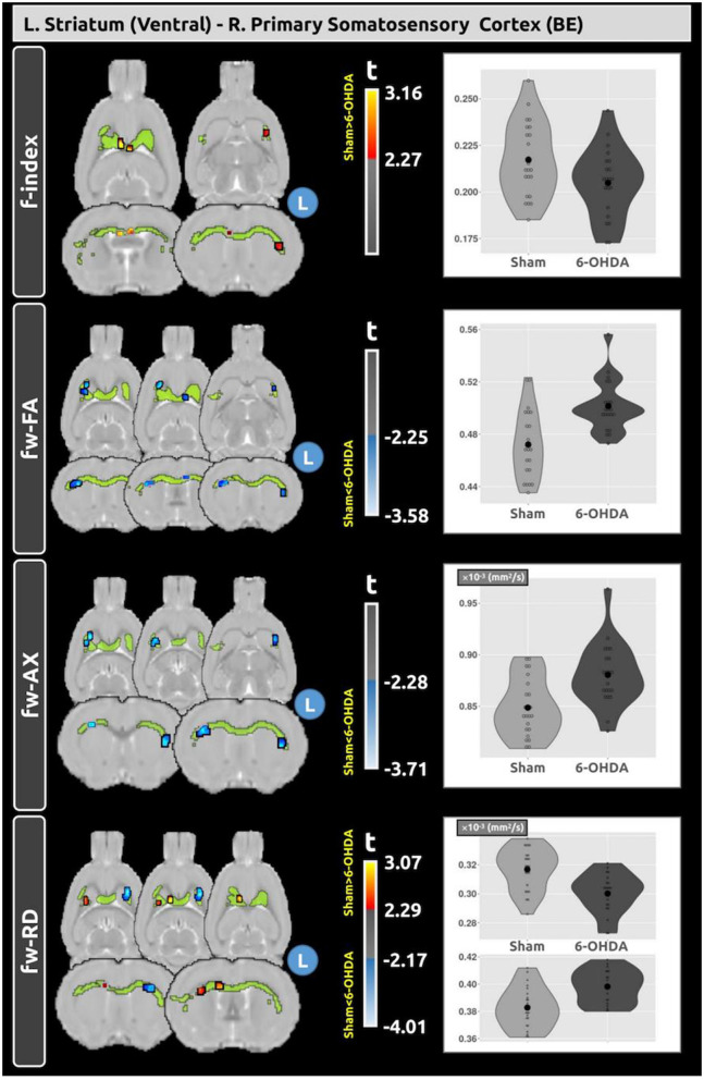
Functionally, decreased connectivity between the retrosplenial and endopiriform cortices in the 6-OHDA model suggests disrupted sensory processing, while increased connectivity between the hippocampus and retrosplenial cortex indicates possible compensatory mechanisms. Structurally, we observed reduced connectivity between the subcoeruleum and piriform cortex in the 6-OHDA model, which may reflect axonal degeneration, and increased connectivity between the ventral striatum and primary somatosensory cortex, which likely reflects compensatory changes to support motor-sensory integration. Diffusion MRI analysis further revealed changes in the white matter tracts connecting these regions, supporting these findings and highlighting adaptive responses to neurodegeneration in PD.
These findings demonstrate the utility of combining functional and structural connectivity analyses to capture PD-related network disruptions. These structural connectivity changes were further associated with microstructural alterations. The development of MRI biomarkers for understanding brain connectivity may enhance our understanding of PD pathology and advancing translation of these techniques to clinical applications.
帕金森病(PD)是第二常见的神经退行性疾病,其特征是与多巴胺能神经元变性相关的运动和非运动症状。本研究利用6-羟基多巴胺(6-OHDA)大鼠模型,通过向内侧前脑束和黑质进行靶向注射,复制类似PD的多巴胺能变性。
行为评估揭示了典型的运动缺陷,同时进行MRI以评估互补的功能连接和结构连接。死后酪氨酸羟化酶(TH)染色证实了广泛的多巴胺能神经元丢失,验证了模型的病理相关性并确保了数据完整性。在7T条件下对46只雄性Fischer F344大鼠(23只6-OHDA组,23只假手术组)采集MRI数据,以表征两组之间的功能和结构连接差异。
在功能上,6-OHDA模型中扣带回后部与内梨状皮质之间的连接性降低表明感觉处理受到破坏,而海马体与扣带回后部皮质之间的连接性增加表明可能存在代偿机制。在结构上,我们观察到6-OHDA模型中蓝斑下核与梨状皮质之间的连接性降低,这可能反映了轴突变性,而腹侧纹状体与初级体感皮质之间的连接性增加,这可能反映了支持运动-感觉整合的代偿性变化。扩散MRI分析进一步揭示了连接这些区域的白质束的变化,支持了这些发现,并突出了对PD神经变性的适应性反应。
这些发现证明了结合功能和结构连接分析以捕捉与PD相关的网络破坏的实用性。这些结构连接变化进一步与微观结构改变相关。用于理解脑连接性的MRI生物标志物的开发可能会增强我们对PD病理学的理解,并推动这些技术向临床应用的转化。